Articles

Dislocated Intraocular Lens

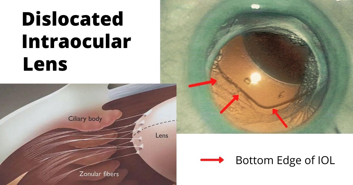
A dislocated intraocular lens is very similar to a dislocated crystalline (natural) lens. The only difference is that the intraocular lens has been inserted during previous cataract surgery.

Cataract surgery is a low-risk surgery and has a success rate of about 95%. But like all surgeries there can be complications, especially if you have other eye or health issues.  

One of those complications is a dislocated intraocular lens (IOL). This is a rare complication and occurs in less than 3% of cases, but it may require additional surgery to repair.

What causes a Dislocated?

Sometimes the dislocation happens shortly after cataract surgery because the zonules, a ring of tiny thread-like fibers that support and hold the lens capsule in place, fail or break. The zonules are made of a connective tissue protein. People who have a connective tissue disorder are more likely to have this complication, and it can also occur in patients who have a history of previous eye surgeries, trauma to the eye, or who take prostate medication.

Other times the IOL dislocation happens gradually over time when the zonules slowly weaken and don’t adequately support the lens capsule and this causes the lens to shift away from the center of the pupil. This happens more often in patients with a history of eye trauma, multiple eye surgeries, especially retinal detachment repair, and complicated original cataract surgery.

Symptoms

The most common symptom of a dislocated IOL is blurry vision. The degree of blurriness depends on how dislocated the IOL is. If the IOL is only slightly dislocated then vision may still be clear, but ghost images or a double image at night may occur when light passes through the edge of the dislocated lens.

Treatments

Treatment depends on the severity of the dislocation. Some dislocations very minor and vision is still good. In these cases, no treatment is necessary.  

If the lens is completely off center and vision is very blurry, then only surgery will correct it. In some cases, the original implanted IOL can be repositioned and secured in place. In other cases when the zonules are extremely weak or damaged, a new IOL designed to be sutured to the wall of the eye (sclera) or to the iris is used.

If the lens has fallen into the vitreous cavity, then a retinal surgeon will need to perform a vitrectomy to remove the vitreous jelly inside the eye and retrieve the dislocated intraocular lens.  

Prognosis

Most patients who have IOL repositioning or IOL replacement have vision of 20/40 or better after surgery. The outcome depends on your health and if you have other eye issues, such as macular degeneration or diabetic retinopathy.  

Recovery for IOL replacement is similar to the recovery time for cataract surgery. The medications are the same and most patients can resume normal activities the day after surgery.

If you would like to schedule an appointment, please call us (877) 245.2020.
Nader Moinfar, M.D., M.P.H.
Retina Specialist
Orlando, FL

4 Week Dosing Beovu Study was Halted

Company halts a new BEOVU study, a drug for wet macular degeneration, as increased inflammation is noted.

A Beovu study was halted recently.

In a May 28, 2021 press release, Novartis Pharmaceuticals halted an ongoing trial of BEOVU (brolucizumab) for the treatment of wet macular degeneration. About 1 year ago, there were concerns over Beovu and potential complications.

BEOVU, an anti-VEGF drug, is FDA approved for the treatment of wet macular degeneration and is delivered via intraocular injection. The approval also allows for the drug to be repeated as often as every 8 weeks. The trial was to determine if Beovu could be administered every 4 weeks instead of 8. Most of the other anti-VEGF medications can be given every 4 weeks.

Other anti-VEGF medications include:

  • Lucentis (ranibizumab)
  • Avastin (bevacizumab)
  • Eylea (aflibercept)

All are given by intraocular injection and are powerful weapons for the treatment of macular degeneration, diabetic retinopathy and retinal vascular occlusions.

Why Study was Halted

This was originally a 2 year study where patients were treated every 4 weeks with Beovu. Great clinical studies are so-called “double blinded”, that is, neither patient nor treating physician knows what drugs are being delivered. This prevents bias in the study.

The results of the first year of the study were recently released which showed, when dosed at a higher frequency (4 weeks), an increase in the incidence of intraocular inflammation when compared to other studies.

The Beovu study was halted in the interests of patient safety. While most intraocular inflammation is well tolerated, that is, there is no long term sequalae, this conservative approach may save vision in many patients involved in the study as some patients may have permanently lost some vision.

This does not mean Beovu is not a good drug, it just means that studies indicate that the drug may have some toxicity if used too often. In the case of Beovu for macular degeneration, use every 4 weeks may be too often.

Presently the FDA has approved the drug to be used no more often than every 8 weeks.

Beovu Potential

Several studies have found that Beovu, when given only every 8 weeks, is a very potent and effective drug for wet macular degeneration, particularly in those patients where other drugs were unable to control the disease.

Ongoing clinical trials are testing the effectiveness of Beovu for treating diabetic macular edema (DME), a complication of diabetic retinopathy. Other anti-VEGF medications are also approved for the treatment of DME.

Retina specialists vary in their preference of anti-VEGF medications for certain conditions. Patient response to a particular medication often varies, for instance, consider the use of Tylenol, aspirin and ibuprofen. Though all can be used for pain relief, most of us find that one works better for us than the others.

It is very likely that Beovu will find it’s place in treating hard to treat patients with wet macular degeneration.

Stay tuned.

If you would like to schedule an appointment, please call us (877) 245.2020.

Nader Moinfar, M.D., M.P.H.
Retina Specialist
Orlando, FL

Dislocation of the Natural Lens

The fancy term for diplacement of your natural lens in the eye is called ectopia lentis…here’s when it happens.

Your natural lens can become displaced from it’s natural anatomic position within the eye. Intraocular lenses can also become displaced, but this article is limited to dislocation of the natural crystalline lens.

Ectopia lentis is the medical term for the dislocation or displacement of the eye’s natural crystalline lens. The lens may be free-floating in the vitreous or it may be in the anterior chamber or directly on the retina.

This dislocation most commonly occurs after trauma to the eye. It can also be caused by a systemic disease, such as Marfan syndrome. Trauma is the most common cause and most often the result of a direct blow to the eye such as from a baseball or golf ball.

Some rare and inherited genetic diseases can cause ectopia lentis:

  • Weill-Marchesani syndrome
  • Sulfite oxidase deficiency
  • Hyperlysinemia
  • Ehlers-Danlos syndrome
  • Sturge-Weber syndrome
  • Mandibulofacial dysostosis
  • Wildervanck syndrome
  • Conradi syndrome
  • Pfaundler syndrome
  • Crouzon syndrome
  • Pierre Robin syndrome
  • Sprengel deformity

Symptoms of Lens Dislocation

The most significant symptom of ectopia lentis is reduced visual acuity such as poor near or distant vision. The degree of reduction in visual acuity varies with the degree of lens dislocation and the type of dislocation.

If the zonules in the eye are disrupted it can lead to increased curvature of the lens and may result in lenticular myopia or astigmatism. The zonules are tiny thread-like fibers that hold the eye’s lens firmly in place and also tighten the pull the lens to accommodate near vision.

Treatment

Treatment of ectopia lentis depends on where and how far the lens has moved and any resulting complications. In some cases, in which the dislocation is minimal and there is no significant impact on vision observation and close follow-up are the only treatments necessary.

In cases in which vision is affected or there is damage to surrounding structures, surgery may be necessary. The dislocated lens is removed, and an artificial lens is put into place. If there is not enough structural support for the artificial lens then it may need to be sutured to the iris or sclera.

Prognosis

Multiple surgical techniques are available for correction of ectopia lentis and each has its own limitations and associated complications. In some cases, an implanted artificial lens is used, and other patients are treated by removal of the lens and a vitrectomy to remove the vitreous humor and then they use special contact lenses.  

If the lens dislocated due to a genetic disease then the underlying disease must also be treated.

Most patients with ectopia lentis do well and at least 85% achieve a 20/40 or better visual acuity. However, if there is a pre-existing condition such as a corneal disease, glaucoma, diabetic retinopathy, macular degeneration, or a history of retinal detachment, the outcome will not be as favorable.

Close follow-up is essential as is medical management of any complications that may arise. 

If you would like to schedule an appointment, please call us (877) 245.2020.

Nader Moinfar, M.D., M.P.H.
Retina Specialist
Orlando, FL

Vitrectomy for Retinal Detachment Repair

This is the most common way to repair most retinal detachments and has faster healing times.

Vitrectomy for retinal detachment repair is an alternative method to scleral buckle or pneumatic retinopexy.

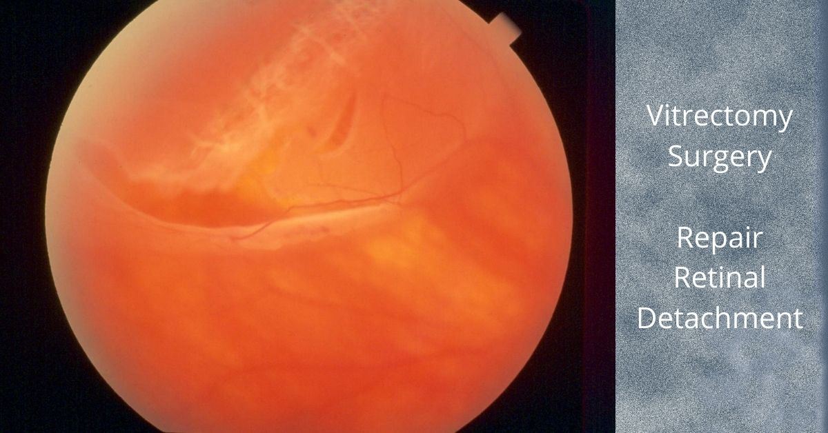
A vitrectomy is a common surgical repair for a detached retina. The surgery removes the vitreous gel from the eye, pushes the detached retina back into place, and seals any retinal tears in the retina. Then gas is injected into the eye to help keep the retina in place and prevent any fluid from leaking from the repaired tears and into the subretinal space.

The Surgery

A vitrectomy is usually performed using local anesthetic. A heavy sedative is administered and then Lidocaine is injected around the eye to numb it. If patients prefer not to be awake during the procedure they can be given intravenous sedation or a general anesthesia.

The surgery consists of making three small needle-sized incisions (about the width of an eyelash) into the white part of the eye (sclera). Through one incision a constant flow of fluid is passed into the eye. The second incision is used to insert a fiber-optic light to provide illumination of the eye, and the third is for any instruments that are used, including a “vitrector” for the removal of the vitreous.

A vitrector is an oscillating microscopic cutter that slowly removes the vitreous. Once the vitreous is removed, a freezing instrument or a laser is used to seal any tears in the retina.

The vitreous will be replaced with a gas. In time the gas will dissipate and be replaced by the eye’s own fluids (aqueous humor). The vitreous gel does not grow back, but the eye can function with just the fluid that replaces the vitreous. Depending on the type of gas used and its concentration, it can take from three to ten weeks to fully dissipate.

Most retina specialists will perform vitrectomy for retinal detachment repair in an outpatient setting. The surgery time ranges from 20 to 40 minutes, but varies greatly.

Head positioning

Head positioning after surgery is necessary to keep the gas bubble in the proper place. If the retina detached at the bottom, the only way to get the bubble to press against the place of detachment is to place the head down. The bubble walls off the damaged area while it heals and prevents any fluid from flowing through the damaged area of the retina and into the subretinal space.

The gas bubble wiur vision while it is in place, but as it dissipates a line will form across your vision where the newly forming fluid (aqueous humor) is gradually replacing the bubble. The line will move lower each day and the field of vision will get larger.

If you have to position your head down there are pillows, chairs, and mirrors that allow you to see around the room while in a face-down position.

Gas bubble precautions

As long as the gas bubble remains in your eye, you must not fly in an aircraft. The reduced pressure in the cabin will cause the gas bubble to expand. This would be extremely painful and could lead to loss of sight. If you must have general anesthesia for any surgical procedure while the gas bubble is in your eye, you must inform your anesthesiologist so you will not be given nitrous oxide, which would cause a dangerous rise in eye pressure in the eye with the gas bubble.

Safety

Vitrectomy surgery was first developed in 1970 and since then many improvements in instrumentation and technique have dramatically improved the safety of the procedure. Severe complications are rare and the surgical success rate is over 90%.  

Nader Moinfar, M.D., M.P.H.
Retina Specialist
Orlando, FL

If you would like to schedule an appointment, please call us (877) 245.2020.
Port Delivery System | Lucentis Reservoir

The Portable Delivery System may reduce the need for repeated injections of Lucentis

Roche Pharmaceutical is testing the Port Delivery System (PDS), a system which may reduce treatment burden by reducing the number of injections of Lucentis used to treat wet macular degeneration.

Ranibizumab (Lucentis) is a valuable FDA approved tool for the retina specialist to treat wet macular degeneration, diabetic macular edema, diabaetic retinopathy, retinal vein occlusions and myopic choroidal neovascularization (mCNV).  

Ranibuzumab is an anti-VEGF medication that is delivered only by intravitreal injection.  Injections often need to be repeated at least monthly for an indefinite period. 

Treatment Burden

Scheduling repeated medical appointments for these injections can be a burden to many patients.  The injections might be uncomfortable (usually not), may require scheduling a companion for transportation and possible complications are possible with each injection. 

The Port Delivery System has been designed to address the issues of treatment burden to include extending time in between treatments, I.e. fewer injections and scheduled appointments. 

How it works

Initially, a tiny reservoir, about the size of a grain of rice, is surgically fixated to an area inside the eye called the pars plana.  The reservoir serves as a refillable source of ranibizumab (Lucentis) and will release the drug slowly over several months.  When empty, the reservoir can be refilled without its removal.  The technique will require a special needle and can be completed in the office.  It does not require actual surgery. 

Port Delivery System

The system is under FDA investigation (Archway phase 3 clinical trial) and is not approved for clinical use.  Most recently, early Phase 3 data from the Archway study indicate the Port Delivery System is comparable in effect to patients receiving monthly intravitreal injections of ranbizumab (Lucentis).  The Port Delivery System is refilled every 24 weeks. 

Sustained release and slow-release systems may solve the treatment burden for patients requiring repeated injections of anti-VEGF medications such as Lucentis.  In addition, investigations are ongoing to find drugs that have an extended treatment period, sustained release medications all to reduce treatment burden, but achieve comparable or superior results. 

If you would like to schedule an appointment, please call us (877) 245.2020.

Nader Moinfar, M.D., M.P.H.
Retina Specialist
Orlando, FL

What is Scleral Buckle Eye Surgery

Scleral buckle eye surgery has been around for generations and is still a mainstay of retinal detachment surgery.

Scleral buckle eye surgery to repair a retinal detachment has been in use since the early 1950s and it remains an essential technique for reattaching the retina.

The scleral buckle, which is usually made of silicone semi-hard plastic, supports the retina from outside the eye. It is placed under the eye muscles and usually encircles the sclera or white part of the eye and eliminates the tugging that is detaching the retina. 

The buckle is placed behind the eyelids and cannot be seen. It is sewn to the sclera (the actual white part of the eye) and it presses the walls of the eye inward, taking pressure off the retina and allowing it to reattach to the eye’s interior wall. The buckle usually remains in place permanently.

Scleral buckling is sometimes a stand-alone treatment for the repair of a retinal detachment and sometimes it is used in combination with a vitrectomy.

A laser or a sub-zero probe (cryopexy) may also be used to seal off the areas of the retina that were torn to prevent further fluid leakage and pressure on the retina.

Side effects of a scleral buckle

The possible complications or side effects of scleral buckling surgery include:

  • Increased myopia (nearsightedness) caused by the increased length of the eye when it is buckled
  • Increase in astigmatism also caused by the buckle changing the eye’s shape
  • Double vision is uncommon but can occur because the eye’s muscles were manipulated when placing the buckle under the eye muscles.
  • Pain after the procedure which can usually be controlled with acetaminophen or ibuprofen and will subside in a few days.

 What causes retinal detachment?

The most common type of retinal detachment, that accounts for 90% of all retinal detachments,occurs when a small tear or hole appears in the retina that allows the vitreous gel of the eye to seep behind the retina where it pushed the retina away from the back of the eye. 

Tears in the retina are caused by vitreous gel, which contains millions of fibers, that becomes “sticky” and tugs at the retina making minute tears in it. Retinal holes develop in areas where the retina has thinned. Retinal holes are typically smaller than tears and have a much lower risk for causing a retinal detachment.

Recovery after scleral buckling eye surgery

The recovery time varies, but ranges from two-to-eight weeks. An eye patch may be required for a few days after surgery and antibiotic eye drops may be prescribed to prevent infection. Your eye may have redness, tenderness, and swelling for a few weeks after surgery.

If you would like to schedule an appointment, please call us (877) 245.2020.

Nader Moinfar, M.D., M.P.H.
Retina Specialist
Orlando, FL

Vitreomacular Traction Syndrome

VMT is very similar to macular pucker and causes the same changes to your vision.

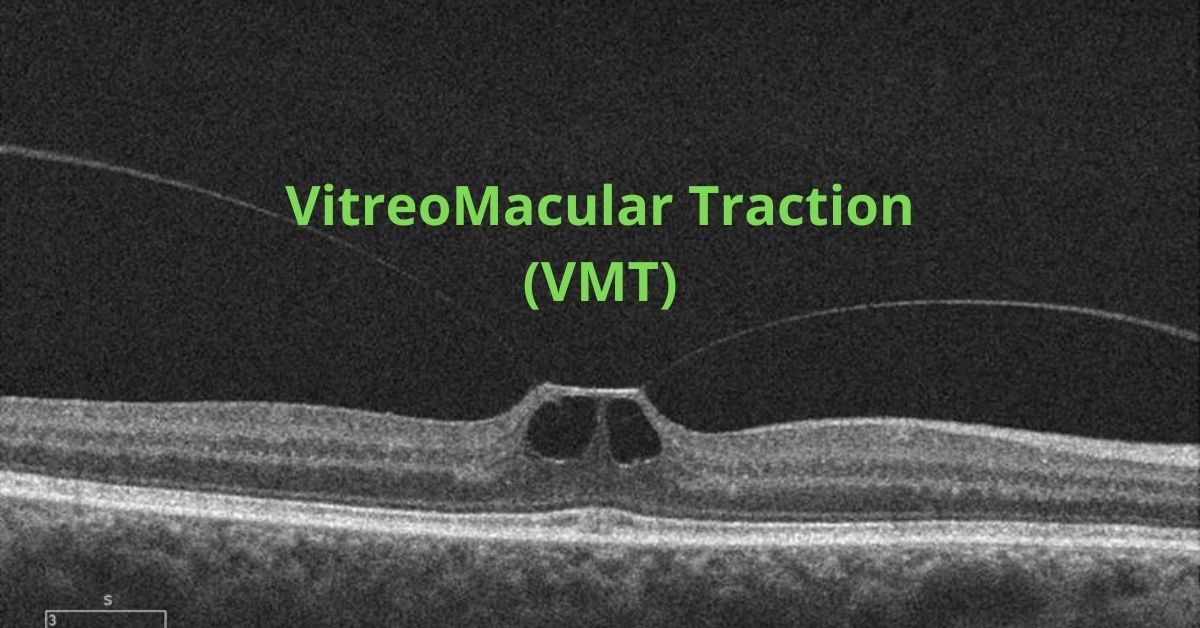
Vitreomacular traction (VMT) is a complication that arises from a normal process. That normal process is posterior vitreous detachment (PVD) and it happens to everyone as they age. By about age 40 or 50 the vitreous gel that fills the eye begins to change. It begins to shrink and lose fluid and strands of the gel can drift through the eye. These strands can be seen as dark strings or spots that float around in your field of view and are called floaters.

The vitreous gel eventually completely separates from the retina. This is perfectly normal and happens to most people by the age of 70, that is, a PVD is a completely normal event.

Problems arise only if the vitreous gel is strongly attached to the retina, specifically at the macula. If that is the case then when the vitreous gel shrinks it can pull on the retina.

That sticking process is called vitreomacular traction and is very similar to an epiretinal membrane.

The pulling and tugging on the center of the retina where the macula is located can damage the macula and cause vision loss if left untreated.

In healthy eyes, VMT is not common. Certain eye conditions or diseases put people at a higher risk of developing VMT. Those conditions and diseases include:

  • High myopia which is extreme nearsightedness
  • Age-related macular degeneration which is a breakdown of the tissues in the back of the eye
  • Diabetic eye disease which affects the blood vessels in the back of the eye
  • Retinal vein occlusion which is a blockage of veins in the retina

Symptoms

The most common symptoms of VMT include:

  • Distorted vision that makes straight lines appear wavy, blurry or have blank spots
  • Seeing lots of flashes of light in your vision
  • Seeing objects as smaller than their actual size

It is important to see an ophthalmologist for an evaluation when you first notice any of these symptoms.

Diagnosis

To diagnose vitreomacular traction (VMT) your ophthalmologist will use tests such as optical coherence tomography (OCT) which uses light waves to take pictures of the various layers of the retina and will show any damage to the macula.

Fluorescein angiography may also be used. This is an imaging test to view how well the blood is circulating inside the retina and to find any macula swelling. It uses a medical imaging dye injected into the arm that circulates inside the retina while a special camera photographs the progress of the dye as it moves through the blood vessels in the eye.

An ultrasound scan may also be used so your ophthalmologist can get a better view of the location of the sticking point between the vitreous and the macula.

Treatment

Some cases do not require treatment and will resolve on their own, but you will be asked to monitor your vision at home with a grid of lines to make sure the VMT does not progress. If the lines on the grid begin to appear wavy or have missing areas then you will most likely require treatment.

Surgery to remove the vitreous and replace it with a saline solution may be needed to prevent macular holes, puckers or macular swelling from developing or worsening.

Some people are candidates for medication treatments. A medication that dissolves the proteins that link the vitreous to the macula can be injected into the eye. Usually only one injection is needed.

Prognosis

Most patients with VMT maintain good visual acuity in the affected eye even if treatment is required.

If you would like to schedule an appointment, please call us (877) 245.2020.
Nader Moinfar, M.D., M.P.H.
Retina Specialist
Orlando, FL

Cystoid Macular Edema

This type of macular swelling is very common and even occurs after cataract surgery.

What is Cystoid Macular Edema?

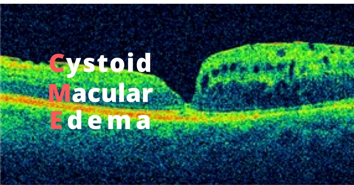
Cystoid macular edema, aka CME, affects the functional center of the retina, the macula.  It is another term for macular edema where multiple cyst-like areas of swelling develop.

There are multiple causes of CME to include:

  • Retinal Vascular Occlusions (RVO)
  • Intraocular inflammation (uveitis or iritis)
  • Diabetic retinopathy
  • Uncomplicated cataract surgery

Symptoms of Cystoid Macular Edema

The main symptom of CME is blurry vision.  There may also be some symptoms of distortion, poor color perception and minification, but most patients note blurry vision.

Because this is a condition of the macula, only central vision is affected.  The peripheral or side vision is not involved.

Blurry vision can occur from a variety of eye conditions and is not a specific symptom of cystoid macular edema. 

CME after Cataract Surgery

About 1-3% of patients will develop cystoid macular edema following cataract surgery.  This can occur several weeks following uncomplicated cataract surgery.  Before modern phacoemulsification techniques evolved, CME developed in as many as 10% of patients.

For reasons unknown, fluid starts to accumulate in the macula.  Patients are often frustrated because the vision following surgery was excellent, only to get worse. 

Treatment and Prognosis

CME following cataract surgery is usually self limited, that is, there is full restoration of vision and function after several weeks of treatment.  The condition does not usually recur, but there is increased risk of developing CME in the fellow eye.

Treatment for CME following cataract surgery often involves topical anti-inflammatory eye drops.  Steroid drops are often quite effective.  In more complicated cases, injections of steroid around the eye may be helpful

Because CME can develop from other situations other than cataract surgery, treatments and prognosis vary widely.  For instance, cystoid macular edema following a branch retinal vein occlusion may involve anti-VEGF injections or depot steroids.  

Cystoid macular edema can also be caused by certain glaucoma drops or following complicated cataract surgery.  Surgery may be necessary to treat the macular swelling due to complications of cataract surgery. 

Consultation with a retinal specialist may be needed.

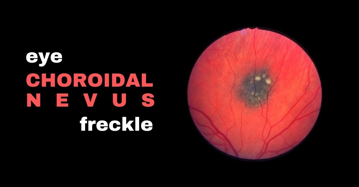
What is a Choroidal Nevus?

A freckle in your is usually benign, but there is a concern about choroidal nevi.

A choroidal nevus is the fancy term for a freckle in the retina.

A nevus is the medical term for mole. Nevi is the plural of nevus. A nevus can appear on your skin, the surface of the eye or inside the eye. 

When a nevus is inside the eye it is called a choroidal nevus because those types are found under the retina in the layer of tissue called the choroid.

A nevus is a cluster of melanocytes—the cells that produce melanin. They are the pigment that colors hair, skin, and eyes. They are much more common in people with lighter skin tones.

They are often referred to as eye freckles and the ones that appear inside the eye are discovered during routine eye exams and are usually harmless and most of them won’t affect your vision or cause any problems and they require no treatment except monitoring the nevus for changes.

Three Types of Eye Nevi

  • A nevus in the white part of your eye, conjunctival nevus, are common and range from yellow to brown and can lighten or darken over time. 
  • Nevi in the iris (the colored part of your eye) are tiny, dark brown flecks on the surface of the iris and can grow larger over time but are usually harmless and usually do not become melanomas. 
  • A choroidal nevus is typically gray, but can be brown, yellow, or variably pigmented. They are also mostly harmless.  

Most nevi do not need to be treated and will not affect your vision or lead to any other health problems. The only reason you might need treatment is if your doctor suspects the nevus might be a melanoma.

What Causes a Nevus?

The cause of eye nevi is not known, but there are associations between eye nevi and UV light. Wearing sunglasses to protect your eyes from ultraviolet light is always recommended.

A Choroida Nevus can be Suspicious for Cancer

A very small percentage of choroidal nevi have features that make them at a higher risk of growing into a melanoma. Those choroidal nevi should be monitored by your doctor through regular eye exams. Very rarely, a choroidal nevus may leak fluid or be linked to abnormal blood vessel growth.

There are five factors that signal the choroidal nevus should be closely monitored:

  • Thickness of greater than 2.00 mm
  • Subretinal fluid
  • Visual symptoms
  • Orange pigment of the nevus
  • A part of the nevus touching the optic nerve

Very often, suspicious cases are referred to a retina specialist or ocular oncologist for further evaluation.

If you would like to schedule an appointment, please call us (877) 245.2020.
Nader Moinfar, M.D., M.P.H.
Retina Specialist
Orlando, FL

Face Mask May Increase Eye Infections

Here’s another reason to be careful about wearing a mask.

Wearing a face mask is important for patients from the moment they walk into a clinic to protect themselves and others from SARS-CoV-2 infection. But additional safety measures should be used when the patient is wearing a face mask and receiving intravitreal injections to prevent contamination that could occur when exhaled breath is blown toward their eyes.

A study to investigate the air that escapes from the top of face masks used a thermal camera and found that air jets originating from the top edges of face masks and radiating toward the eye were detected in 81% of cases.

3 Face Masks

Three face masks were tested. A regular face mask with four tying strips, one with elastic ear loops, and the 220 N95 tuberculosis particulate face mask.

Exhaled breath that is normally blown forward is blocked by face masks and that air is redirected toward the eye. People who wear glasses and face masks are aware of this issue. It causes their glasses to fog up.

Anti-VEGF injections have a very low risk of endophthalmitis. Several large studies have estimated the risk at approximately 0.05% or 5 in 10,000 for any individual patient receiving monthly injections. Having face masks blowing air into the eye could increase that risk slightly and for that reason extra precautions are recommended.  

Study Recommendations

The additional precaution can be done in one of two ways—a surgical drape can be used to isolate the eye or medical adhesive tape can be applied on the upper border of the face mask to block air passage. The study also recommends using an eyelid speculum to keep the eye open and prevent the eyelid from coming into contact with the needle.

The study’s author, Amir Hadayer, MD, says, “Protocols for preparing patients for intravitreal injections vary and not all specialists use draping and a speculum in normal times, but under the current circumstances, these extra precautions are recommended.”

What is Endophthalmitis?

Endophthalmitis is an infection that causes severe inflammation of the tissues inside the eye. The infection is typically caused by bacteria for fungi.

Symptoms of Endophthalmitis

  • Decreased vision
  • Pain
  • Redness or swelling of the eye

These symptoms don’t always mean you have endophthalmitis, but if you experience one or more of these symptoms, contact your ophthalmologist.

If you would like to schedule an appointment, please call us (877) 245.2020.
Nader Moinfar, M.D., M.P.H.
Retina Specialist
Orlando, FL

Jon Doe