Articles

Dilated Eye Exams

Why you need to have your eyes dilated by a retina specialist.

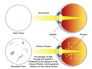
In order to properly examine your retina, a dilated eye exam is necessary each and every time you come to the office. A dilated eye exam requires use of dilating drops to dilate and enlarge your pupils to allow us to properly diagnose and treat diseases of the retina.

A dilated eye exam is the only way the retina can be examined by direct visualization using a combination of lenses and specialized ophthalmic instruments.

Other methods such as photography, fluorescein angiography, optical coherence tomography (OCT) and ultrasound can be used in conjunction to direct visualization to diagnose and to treat retinal diseases.

As a retina specialist, virtually everyone of my patients needs a dilated eye exam every time they come to the office.

Dilating Eye Drops

There are several drops used routinely to dilate the eyes. All are marked with a bright red cap to distinguish these eye drops from other types.

In most cases, we only need to use phenylephrine and tropicamide do satisfactorily dilate the pupils. These drops work differently to achieve dilation.

Tropicamide inhibits the small muscle located at the margin of the pupil. The function of the pupillary sphincter muscle is to constrict the iris in bright light and makes the pupil smaller through a purse-string mechanism. Tropicamide prevents this constriction.

Phenylephrine stimulates another set of muscles which function to enlarge the pupil. The iris dilator muscle runs radially along the iris and, when stimulated, pulls the iris open.

Cyclopentolate and atropine can also dilate the pupils but their effects last much longer (days to weeks) and are generally not used for diagnostic purposes.

How Long does Dilation Last?

In most circumstances, your eyes will stay dilated for several hours after the exam. Several factors influence the length of time your eyes will stay dilated, such as:

  • Color of your eyes
  • Your age
  • Type, strength and amount of the medicine
  • Previous eye surgery

Lightly colored eyes, such as blue and hazel, dilate very quickly and stay dilated longer compared to brown eyes.

Brown eyes actually contain brown colored pigment which absorbs the dilating medicine, whereas blue eyes actually contain no pigment and the eyes dilate faster.

Younger eyes tend to take a longer time to dilate. Certain diseases (e.g. iritis) can make dilation take a very long time.

Dilation usually takes at least 20 minutes at a minimum and will reverse after a few hours.

If you would like to schedule an appointment, please call us (877) 245.2020.

Nader Moinfar, M.D., M.P.H.
Retina Specialist
Orlando, FL

Beovu | New Treatment ARMD

This new FDA approved drug for wet macular degeneration requires fewer injections.

The FDA recently approved another intravitreal injection for the treatment of wet-ARMD. Beovu (brolucizumab) is an anti-VEGF medication which may be useful for extending the frequency of injections in patients requiring multiple treatments.

The FDA found that Beovu may be useful in the treatment wet macular degeneration and offers an extended 3 month dosing schedule.

Anti-VEGF Medications

Other anti-VEGF medications for the treatment of wet macular degeneration include:

  • Avastin
  • Lucentis
  • Eylea
  • Macugen

Wet macular Degneration

There are two forms of macular degeneration: wet and dry. The dry form accounts for about 90% of patients with macular degeneration. Symtoms of distortion, blind spots and blurry vision are less so than the “wet” counterpart.

Wet macular degeneration accounts for the other 10% of ARMD patients. Wet macular degeneration is more aggressive and causes more pronounced and quicker loss of vision.

Wet macular degeneration occurs with the development of abnormal blood vessels growing within the layers of the retina. The abnormal blood vessels, aka neovascularization, are stimulated by a protein called Vascular Endothelial Growth Factor (VEGF).

anti-VEGF medications, like Beovu, work by neutralizing the VEGF protein making it ineffective. As a result, the neovascularization regresses and can not be stimulated to grow.

Timing of Injections

All the anti-VEGF medications work in similar fashion and share similar results. In addition, the dosing schedule of all the anti-VEGF injections usually ranges between every 4-6 week dosing (a shot every 4-6 weeks).

Beovu was compared to Eylea to gain FDA approval. Beovu may have the advantage of achieving similar results (non-inferior) to Eylea, but may be given once every 3 months compared to monthly injections.

FDA approval usually only gives the retina specialist guidance or suggestions on a drug’s use. Though dosing is approved for every 3 months, it does not necessarily mean that a retina specialist will find this necessarily true in practice.

Most retina specialists will slowly try the new drug and determine for ourselves which type of patients will derive the most benefit in terms of treatment efficacy and dosing.

If you would like to schedule an appointment, please call us (877) 245.2020.

Nader Moinfar, M.D., M.P.H.
Retina Specialist
Orlando, FL

Retinal Vascular Occlusions (RVO)

Retinal vascular occlusions cause painless loss of vision. Here’s a short review of the 4 different types.

There are four types of retinal vascular occlusions (RVO).  All have similar symptoms of acute (sudden) and painless loss of vision.  

In general, vein obstructions occur when the vessel is compressed from the outside of the blood vessel whereas artery obstructions occur when cholesterol plaque (or other substances) travel through the blood stream and become lodged in a blood vessel.

Vein and artery occlusions have characteristic appearances and can be easily diagnosed by a retina specialist.

Artery occlusions are more likely to be associated with coronary artery disease and stroke.

The four types of retinal vascular occlusions include:

  • Branch retinal vein occlusion (BRVO)
  • Central retinal vein occlusion (CRVO)
  • Branch retinal artery occlusion (BRAO)
  • Central retinal artery occlusion (CRAO)

Branch Retinal Vein Occlusion

Compared to a central vein occlusion, this is a “partial” occlusion of the retinal vein.  Usually branch occlusions involve about 50% or less of the retina. If the macula becomes involved, there is usually moderate vision loss.  On occasion, the macula is not involved and vision remains normal. 

Treatment of BRVO includes laser surgery, injections of anti-VEGF and/or intraocular steroids including Ozurdex, a sustained release device which allows continuous treatment for up to 4-6 months.

Central Retinal Vein Occlusion

The entire retinal vein is occluded.  Severe vision loss is normally experienced.  Unlike the branch vein occlusion, the macula is always involved.

Treatment of CRVO may include laser, anti-VEGF injections or sustained release steroids.  Vision usually does not improve as well as the branch retinal vein occlusion.

Branch Retinal Artery Occlusions

Branch artery occlusions do not involve the entire retina and, similar to venous occlusions, less than 50% of the retina is involved.  If the macular area is involved, central vision is decreased. 

Branch artery occlusions do cause retinal edema, but the vision loss is usually more permanent though there is resolution of the retinal swelling.

Both branch and central artery occlusions are caused by an embolus:  usually a cholesterol plaque carried through the bloodstream and becomes lodged in a retinal artery. 

Central Retinal Artery Occlusion

As with the branch artery occlusion, an embolus lodges in the central artery thus cutting off blood flow to the entire retina.  Vision loss is severe and usually does not respond well to treatment.

Historically, many different methods have been described to dislodge the embolus, but none with resounding success.

Patients with artery occlusions should be assessed for the risk of cardiovascular disease and stroke.  It’s important that your retina specialist or eye doctor coordinate care with your primary care physician. 

If you would like to schedule an appointment, please call us (877) 245.2020.

Nader Moinfar, M.D., M.P.H.
Retina Specialist
Orlando, FL

Diseases of the Macula | Distortion

Here’s a short list of retinal diseases that cause metamorphopsia….aka distortion.

Distortion in the vision is usually caused by a disease of the macula. The macula is the functional center of the vision. It is a small area of the retina, yet it yields the most sensitive visual acuity (i.e. 20/20) and our best color perception.

Any change in this tiny area can yield visual distortion: e.g. the perception that objects are crooked, yet we know they are straight. An example is a line of text or the straightness of a telephone pole

Causes of Distortion

There are many macular diseases that may distort the vision. Any disease that causes swelling of the macula or physically changes the macula can cause distortion.

Here’s a short list of a variety of diseases evaluated and treated by a retina specialist:

Diabetic Macular Edema – a common complication of diabetic retinopathy. The macula actually swells due to leaky blood vessels affected by diabetes.

Epiretinal Membrane – also known as macular pucker or cellophane maculpathy. A membrane actually develops on the surface of the retina causing physical distortion of the retinal surface.

Macular Hole – a distinct “stretch hole” develops in the center of the macula causing blind spots and distortion.

Macular Degeneration – either form, wet or dry, can cause distortion.

Retinal Vascular Occlusions – these include retinal vein occlusions and retinal artery occlusions. Macular edema often is associated with occlusion which may cause distortion.

Evaluation of Distortion

I usually recommend that anyone who notices a persistent change in vision, including distortion, call their eye doctor. Problems with the retina usually don’t wax and wane, thus, they don’t cause temporary or fleeting symptoms.

Any one who has complaints of distortion may need to have:

  • Dilated Eye Exam
  • Fluorescein Angiography
  • OCT – Optical Coherence Tomography

Treatment of Distortion

Treatment options are geared toward repairing the physical disruption of the retina. Vitrectomy eye surgery is indicated as in the cases of macular holes and epiretinal membranes. Macular swelling can be treated with a variety of non-surgical methods including drops, injections or laser.

If you would like to schedule an appointment, please call us (877) 245.2020.

Nader Moinfar, M.D., M.P.H.
Retina Specialist
Orlando, FL

Laser Eye Surgery for the Retina

Lasers are commonly used in all of ophthalmology and are useful tools to treat disease of the cornea, glaucoma, cataracts and various retinal diseases.

Because the cornea is transparent to light, the cornea allows the laser energy enter the eye and treat the inner parts of the eye such as the retina.

Lasers for the Cornea

Reshaping the cornea allows many patients to have refractive surgery. Modern refractive surgery is called LASIK. The precision of the laser allows the surgeon to accurately resculp the curvature of the cornea to match the prescription otherwise needed by wearing glasses or contact lenses. An eximer laser is commonly used to perform LASIK.

Laser for Glaucoma

Laser surgery for glaucoma has been long been a treatment option to reduce intraocular pressure. Reducing the internal eye pressure is the mainstay of most treatments for glaucoma. Argon, diode or Neodymium:YAG lasers can be used for glaucoma laser surgery.

Cataract Laser Surgery

There are two ways lasers can assist the eye surgeon to treat cataracts and so-called after cataracts. Laser assisted cataract surgery allows the eye surgeon to more accurately perform several steps of the cataract surgery, but not the entire surgery. A femto-second laser is used for laser-assisted cataract surgery.

After initial cataract surgery, the intraocular lens commonly becomes cloudy. This cloudiness can be improved with a Nd:Yag laser.

Retina Laser Eye Surgery

Lasers are used by a retina specialist to treat 3 different types of problems:

A retinal tear can happen to anyone. Retinal tears can cause a retinal detachment which requires surgery and is vision threatening. Using an argon or diode laser, laser burns surround the retinal tear to prevent a retinal detachment from occurring from that specific tear. In essence, the retinal burn, “seals” the tear, preventing retinal detachment.

Certain types of retinal vascular occlusions (RVO) can cause swelling, aka edema, in the retina. Similar types of swelling are commonly treated in patients with diabetes. The laser acts to decrease swelling by either reducing the rate of fluid accumulation or increasing the rate of absorption.

Advanced retinal diseases, called retinopathy, can be treated with laser to stabilize the eye and prevent vision loss. Proliferative diabetic retinopathy is often treated with laser or intravitreal injections. Retinopathy can also be caused by types of vascular occlusions.

If you would like to schedule an appointment, please call us (877) 245.2020.

Nader Moinfar, M.D., M.P.H.
Retina Specialist
Orlando, FL

Flashes and Floaters

Here’s what you need to know about those pesky floaters and why they could blind you.

Eye “floaters” are deposits or debris within your eye’s vitreous humor. Whether you see specks, wispy threads, strings, or cobweb shapes that drift back and forth when you move your eyes, you are seeing floaters. More precisely, you are seeing the shadows that the floaters cast on your retinas. Floaters can refract light and that temporarily interferes with your vision until the floaters drift into a different position.  

Harmless Floaters?

Eye floaters move around, come and go, and eventually settle away from the line of sight after a few weeks. They can be annoying and distracting, but most of the time they are harmless and are not a cause for alarm, however, any sudden onset of new floaters should be examined.

Floaters are suspended in the vitreous humor which fills about 80 percent of the eye. The most common cause of floaters is shrinkage of the vitreous humor. The vitreous humor is 98 to 99% water and the remaining 1 to 2% is a mixture of collagen, proteins, salts, and sugars. Despite the water-to-collagen ratio, the vitreous humor has a firm jelly-like consistency.

Posterior Vitreous Detachment

As we age the vitreous humor shrinks and liquefies and develops a watery center. Portions of the collagen and protein mixture become stringy fibers and these floating stringy collagen and protein fibers are the floaters. This is called a PVD (posterior vitreous detachment).

Sometimes the small floating vitreous fibers pull on retinal cells and that mechanically stimulates the retina cells causing the sensation of flashes of light. This too is not unusual. Floaters and flashes occur at the same time and are mostly harmless annoyances. You have experienced mechanical stimulation of retinal cells if you’ve ever been hit in the eye and afterwards “see stars”.  

Treatments

In rare instances floaters can be so closely packed and large or so numerous that they significantly affect vision. In those cases, floaters can be treated with a surgical vitrectomy or a laser vitrectomy.  A surgical vitrectomy removes the vitreous humor and replaces it with saline or a bubble made of gas or silicone oil. A laser vitrectomy breaks apart large floaters and vaporizes them.

Floaters and flashes of light that require immediate attention

A sudden onset of numerous floaters, frequent flashes of light, a dark shadow that covers part of your side vision, or eyes that hurt can be symptoms of a tear in your retina and could indicate that your retina is detaching. A detached retina is an emergency and immediate treatment can save your sight. 

If you want to make an appointment, please call us (877) 245.2020.

Nader Moinfar, M.D., M.P.H.
Retina Specialist
Orlando, FL

Eye Injuries

Doing fireworks with my kids last night, reminded me that the forth of July is one of the most common holidays for eye injuries.
Patients can frequently receive burns to the cornea from sparklers. These burns, sometimes also called abrasions, are very painful. The cornea is one of the most highly innervated tissues in the body, and very sensitive to any injury. If the abrasion is small, simple lubrication may be all that is necessary. If larger, then sometimes the cornea may need to be patched.
Not infrequently, a piece of rust or debris may also get embedded in the cornea. In most cases, these pieces lie deeper than they appear, and need to be removed in the doctor’s office.
I’ve operated on several patients who have received an explosive blast injury to the eye, from a bottle rocket, or a fire cracker. In these cases, the eye (also called the globe) is torn open, and the injury is referred to as a ruptured globe; you can read more about ruptured globes under the trauma section of this web site. These injuries tend to be relatively complicated, because the eye becomes very disorganized, and it can be difficult to suture all the pieces back together. Not infrequently, such a patient will need the care of a retina specialist to fix associated problems, like a hemorrhage in the eye, retinal detachment, or foreign body.
Fortunately, most firework-related injuries are preventable. Keeping young children a safe distance from the excitement, and handlers wearing protective eye wear, can help everyone enjoy the festivities with peace of mind.

Treatment of Floaters

“Floaters” is a very broad term, describing objects that seem to float in your vision. By far and away, floaters are not dangerous nor sight-threatening– they are simply changes in the jelly of the eye, called the vitreous, that occurs as we grow older.
In some instances, floaters can represent blood or pigment, that may arise from a retinal tear; you can read more about this under the “retinal tears” section of NaderMoinfarMD.com.
Assuming that the floaters are not from a retinal tear, inflammation or certain cancers of the eye, it is perfectly fine to just leave floaters alone. Over time, much of the visual disturbance will simply diminish, as the floater breaks up and becomes smaller.
I personally have had floaters in both eyes since college, and really don’t even notice them very much. Like most people, I will notice them if I’m reading, or observing something against a white background.
Occasionally, a patient may be referred to me for floaters that are truly interfering with their activities of daily living– pilots, professional drivers, etc. Typically, persons such as these will have a large opacity that is pretty much stuck in their central vision, causing a blind spot– perhaps posing a danger to themselves, and well as others.
Treatment for floaters is widely discussed, and there are really no great clinical studies or randomized trials to suggest what works best. You may read some who advocate doing laser, and some who even offer nutritional advice.
I would suggest that if you are concerned, that you seek the help of a fellowship-trained retina specialist. An experienced retina surgeon can offer the most definite treatment, which is to physically remove the floaters through a procedure called a vitrectomy. You can view videos of how a vitrectomy is performed under the “videos” section of NaderMoinfarMD.com.
Briefly, a vitrectomy is an outpatient procedure performed under local anesthesia. The vitreous is removed, and temporarily replaced with saline; over time, your eye will replace the saline with its own fluid. Recovery time is just a day or two, and patients can usually resume most of their regular activities soon thereafter. If performed correctly, once the floaters/junk are removed, they should not come back.

Treatment of a Retinal Detachment

Retinal Detachment | Nader Moinfar MD | Lake Mary Orlando| Retina Specialist
A total retinal detachment.

A retinal detachment usually when there is pulling on the retina, which causes a tear to occur. Causes of retinal tears are multiple, and include trauma, genetic predisposition, myopia (near-sightedness), to name a few.

A retinal tear should be evaluated by a trained retina specialist, and can usually, and successfully, be simply treated in the office.

A retinal detachment is a more complicated problem, and a well- trained retina specialist can offer you different ways of treating this potentially blinding condition.

In some cases, an office-based procedure called a pneumatic retinopexy can be offered; with appropriate selection of the patient, success rates of up to 80% can be achieved. In the office, a gas bubble is injected into the eye; and either the same day, or maybe a few days laser, laser or cryotherapy is applied. Your retina specialist should discuss with you his or her experience level with this procedure, success rates, and experience levels with the other options, that may be available.

Another method to treat retinal detachments, as seen under the video section of NaderMoinfarMD.com, is a procedure called a vitrectomy. Generally speaking, the vitreous, which is the clear jelly of the eye, is the cause of a retinal tear and traction. By removing the vitreous, one removes the underlying cause. A vitrectomy for a retinal detachment must be careful and completely done, to ensure the greatest chance of success. A seen in my videos, the vitreous is removed, and following a procedure termed an air-fluid exchange, laser is applied around the tears, and finally, the space is filled with a gas bubble.

Lastly, your retina specialist may advise alone, or in combination with a vitrectomy, a procedure termed a scleral buckle; this, too, can be viewed on my video section. With this procedure, a very thin band of silicone is secured on the outside, white part of the eye, helping to relieve the internal traction on the retina; cryotherapy, and a gas bubble, are also used.

With almost all cases of retinal detachments, you will be required to keep your head and body in a certain configuration for a short time; this allows the gas inside your eye to push up against the detachment, allowing it to heal, or “stick’ down. Please note that when the gas bubble is in the eye, you should not travel by air, go up to certain altitudes, and must avoid certain types of general anesthetic inhalation agents if having surgery. Your retina specialist should provide you with a green wrist band to remind you, and others, of this precaution.

If you want to make an appointment, please call us (877) 245.2020.

Nader Moinfar, M.D.
Retina Specialist
Orlando, FL

Vitrectomy for Vitreous Hemorrhage

As you have probably noticed from the videos on NaderMoinfarMD.com, a vitrectomy is a procedure that is used to address many different problems of the back of the eye.
With various modifications, a vitrectomy can be used to treat a retinal detachment, macular pucker/membrane, macular hole, and bleeding inside of the eye– also referred to as a vitreous hemorrhage,
A vitreous hemorrhage implies that the back portion of the eye contains free floating blood. Many conditions can cause a vitreous hemorrhage, but perhaps the most common is diabetes.
Diabetes can affect the eyes in a number of different ways, but more advanced stages, referred to as proliferative diabetic retinopathy, are more prone to cause visual problems. In this condition, abnormal blood vessels start to grow or “proliferate” on the surface of the retina; these vessels are abnormal, very fragile, and bleed easily.
If the bleeding is mild, then a trained retina specialist may elect to perform laser surgery in the office, if not already done.
Sometimes, a retina specialist may also choose to inject various medications into the eye, that can slow, or halt, the proliferation of new blood vessels.
If the blood doesn’t clear, then the option of removing it with vitrectomy surgery can be entertained.
As you can see from the videos on NadrMoinfarMD.com, the set up for this procedure is very familiar. The goals are to safely remove the blood, treat and remove any scar tissue, any apply laser treatment.
As with any other vitrectomy, the patient receives some mild sedative, and an anesthetic injection around the eye.
The area around the eye is then cleansed, and sterile drapes are placed around the eye.
The remaining steps are demonstrated for you on NaderMoinfarMD.com, under the “videos” pull-down menu: three cannulas are secured into the eye, the blood is removed, the vitreous is meticulously cleared, and laser is applied.
A patch is applied on the eye, and patients usually go home 30 minutes or so, after surgery.

Jon Doe