Elective Surgery May Stop

Here’s why your follow up visit or even your surgery may be canceled.

As the COVID-19 pandemic continues to spread, the American Academy of Ophthalmology (AAO), the Centers for Disease Control and Prevention (CDC) and the American Hospital Association have issued recommendations aimed at reducing exposure and preserving healthcare resources, aka, “flattening the curve.”

Will this affect all eye doctors?  Will this affect you?

Stop Routine Care

The American Academy of Ophthalmology is recommending that eye doctors stop routine eye care at this time and focus only on those patients with urgent and emergent conditions.  Examples of “routine” care might include glasses prescriptions, routine follow-up or cataract evaluations. 

Please check with your own doctor if you need to be seen.

As a retina specialist, I may not necessarily “need” to see a patient I see every 6 months with diabetes who is not noticing any changes since the last visit, but I may need to evaluate someone who recently lost vision or developed pain.  

By limiting the number of patients who come through our offices, we are protecting our patients, our staff and ourselves from exposure to the virus.  It is our way of increasing the likelihood that a patient may remain in place and limit the spread of this disease. 

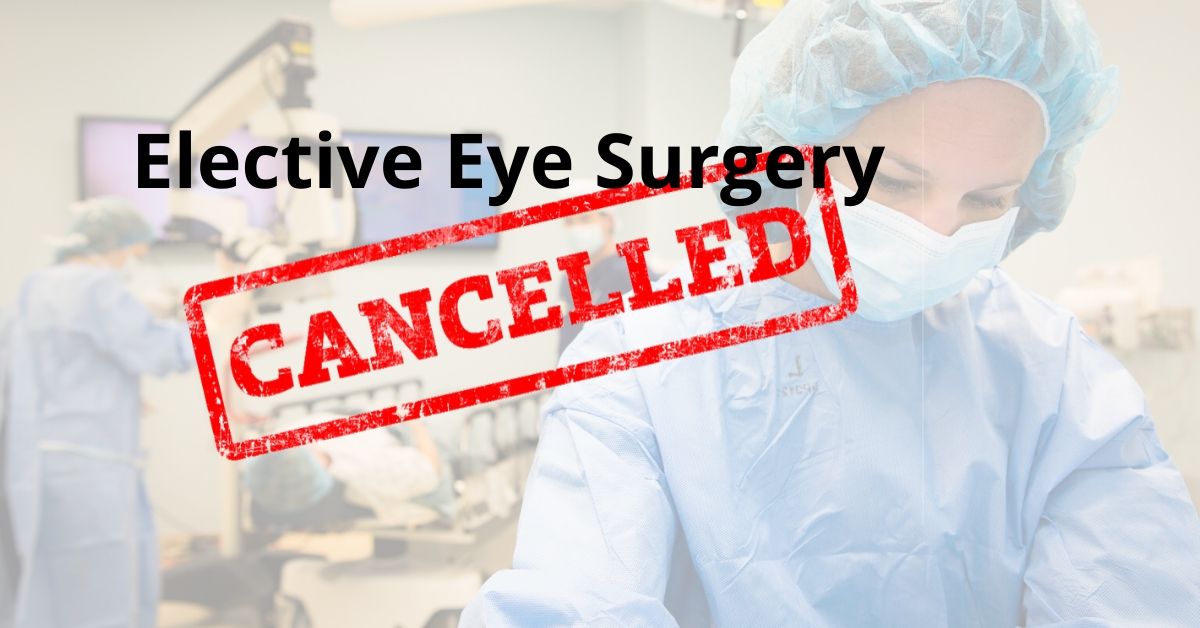
Stop Elective Surgery

Both the American Hospital Association and the Centers for Disease Control and Prevention are recommending that hospitals stop performing elective surgery and non-urgent surgical procedures.  Examples of each might include joint replacements and cataract surgery. Non-urgent surgical procedures might include a colonoscopy.

What’s the rationale?  It saves resources.  

Elective surgeries and procedures take up hospital resources.   By canceling or postponing these procedures, for example, hospital beds and staff can be freed to help with more critical patients elsewhere in the hospital system.

Nurses and aides who normally work in an elective surgical center may be needed elsewhere such as the emergency room.

This practice will also limit exposure to healthy patients.  

Don’t forget that while this pandemic is progressing, patients will still need emergency surgery, chemotherapy, and require care for heart attacks and strokes. 

This is an effort to avoid overburdening our hospital resources and “flatten the curve.”

If you would like to schedule an appointment, please call us (877) 245.2020.

Nader Moinfar, M.D., M.P.H.
Retina Specialist
Orlando, FL

Retinal Disease Urgencies & Emergencies

As the pandemic looms, do you know what urgent and emergent eye conditions might mean?

Eye doctors will likely be asked to see only patients with urgent and emergent conditions. Retinal diseases often require emergent or urgent treatment.

Suggestions have been made to limit office visits to those patients with only urgent and emergency conditions.  In addition, hospitals have been canceling elective surgery and procedures to a later date. Both measures are intended to protect healthy individuals from unnecessary exposure to the virus and to preserve healthcare resources.

Retinal specialists treat “urgent” and “emergent” on a daily basis.  Here’s a short list of what a retina specialist treats on a daily basis.  

Disclaimer:  This article reflects my opinion only.  Your own doctor may have a different opinion.  Please check with your doctor if you have questions or if you are having problems with your vision.  In my opinion, any condition which causes a change in vision or pain should warrant an urgent visit to your doctor….call your doctor.  

N. Moinfar, M.D.

Diabetic Retinopathy

Diabetic retinopathy is the disease diabetes causes in the eye.  Most patients will develop some degree of diabetic retinopathy in their lifetime, and because of this, patients need to be seen regularly whether there are changes in their vision or not.

If there are no acute changes in vision since last visit, then there is probably no urgency to get an examination, though this may not be true in patients with proliferative diabetic retinopathy.  You may call your doctor and discuss how long you can wait after the pandemic is over. 

Diabetes can cause sudden loss of vision due to a vitreous hemorrhage.  Your doctor may want to see you quickly.

New Onset Floaters

Usually new onset floaters are due to a posterior vitreous detachment (PVD).  A PVD can cause a retinal tear which can be diagnosed only by prompt examination and, therefore, new onset floaters should be deemed urgent.  Retinal tears can cause a retinal detachment, hence, the concern.

Macular Degeneration

Patients with macular degeneration can develop loss of central vision and/or distortion.  Many patients self monitor at home with the aid of an Amsler grid, a simple way to look for new distortion in their vision.  If you notice new loss of vision and/or worsening distortion, especially if you are receiving anit-VEGF injections, I would recommend you call your doctor for an urgent evaluation.

Complications from Cataract Surgery

Complications from cataract surgery are usually seen by a retina specialist within a few days of occurrence.  Since cataract surgery is deemed elective, there should not be any complications during the pandemic as cataract surgery is being rescheduled to preserve hospital resources and decrease exposure of healthy people to the virus. 

Retinal Detachment

Retinal detachments can be an emergency, especially if there is progressive loss of vision, but the central vision is spared.  The goal of retinal detachment surgery is to diagnose and perform surgery before the central vision is affected.  

If you would like to schedule an appointment, please call us (877) 245.2020.

Nader Moinfar, M.D., M.P.H.
Retina Specialist
Orlando, FL

Warnings Issued About Beovu

Beovu is FDA approved for the treatment of wet ARMD. An unusual complication may be associated with the drug…

Beovu was recently cleared by the FDA for the treatment of wet macular degeneration. Warnings about its use were recently issued by the American Society of Retina Specialists (ASRS).

Beovu is yet another anti-VEGF medication and, like the others, is delivered directly into the eye by injection. The drugs usually need to be repeated every 4-6 weeks and for an undetermined period.

At the recent meeting of the American Society of Retina Specialists (ASRS), a warning letter was issued to its retina specialist members that a very unusual side effect has been reported.

14 patients with retinal vasculitis have been reported. 11 of the 14 cases were reported to have occlusive retinal vasculitis resulting in severe vision loss. Retinal vasculitis is an unusual complication of any of the anti-VEGF medications.

Anti-VEGF Medications

Other medications used for the treatment of wet macular degeneration include:

  • Macugen (pegaptanib)
  • Avastin (bevacizumab)
  • Lucentis (ranibizumab)
  • Eylea (aflibercept)

Anti-VEGF medications temporarily halt the abnormal growth of neovascularization (abnormal blood vessels) within the layers of the retina. Because the vessels tend to grow back, injections need to be repeated.

Until Beovu was FDA approved, most anti-VEGF injections are repeated every 4-6 weeks. Beovu is unique in that it was approved for a possible 3 month dosing schedule (i.e. an interval of 3 months vs. 4-6 week).

Visual results were comparable to the visual results obtained by Eylea. The drug was not tested against the other anti-VEGF injections in the data submitted for FDA approval.

Novartis Response

Novartis Pharmaceuticals is the manufacturer of Beovu and in two public statements stands by the safety and efficacy of the drug as reported to the FDA.

The company states that the 14 cases of vasculitis fall well within the reported incidence of severe vision loss although it is not clear that vasculitis, specifically, was ever reported as a side effect.

The company is moving forward with its own investigation of these “post-marketing” events and is engaging the help of an external Safety Review Committee (SRC).

If you would like to schedule an appointment, please call us (877) 245.2020.

Nader Moinfar, M.D., M.P.H.
Retina Specialist
Orlando, FL

Macular Degeneration Awareness

February is Macular Degeneration and Low Vision Awareness month. Do you know what other diseases cause low vision?

February is Macular Degeneration and Low Vision Awareness month.  The purpose is to heighten awareness about macular degeneration (ARMD), a leading cause of blindness and low vision.  Prevent Blindness and Bausch & Lomb have again partnered to promote this special month.

Macular degeneration, or ARMD (age related macular degeneration), affects the functional center of the retina, the macula.  The macula provides our sharpest vision and is the only part of the retina capable of providing 20/20 vision. The macula provides us with the ability to read and discriminate things in fine detail.  It also provides us with color vision whereas the peripheral retina only “sees” in shades of grey.

Low vision is defined as loss of vision from eye disease where the vision of the better eye is worse than 20/70.  Common eye diseases include macular degeneration, diabetic retinopathy, congenital/inherited retinal disease (e.g. retinitis pigmentosa), glaucoma, etc. 

Wet and Dry ARMD

There are two types of macular degeneration:  wet and dry. Most patients with macular degeneration have the dry type.  While both types usually affect both eyes, the dry form progresses much more slowly compared to the wet.  There is no cure for either the wet or dry form, though treatments for the wet are available. 

Treatments of the wet form involve injections of anti-VEGF medications.  Oral supplements of AREDS2 vitamins may reduce the chance of severe vision loss in appropriate patients who demonstrate particular risk factors.  Not all patients with ARMD will benefit from AREDS2 supplements.

The implantable miniature telescope (IMT) is also available for select individuals who have significant visual loss from macular degeneration.  The telescope is manufactured by Vision Care and distributed through the CentraSight program.  

Diagnosis of Macular Degeneration

A dilated eye exam is essential in diagnosing macular degeneration.  Patients with ARMD have characteristic findings and your eye doctor will care full examine your retina looking for drusen, pigment changes, fluid and/or blood within the macular area.  There is no specific definitive finding of ARMD.  

Diagnostic testing may involve optical coherence tomography (OCT) and fluorescein angiography (FA) to help make a definitive diagnosis.  

Your eye doctor may consider referral to a retina specialist for further evaluation and/or treatment. 

Other Causes of Low Vision

Diabetic retinopathy and glaucoma are other causes of low vision and are diagnosed and managed differently than macular degeneration.

Other testing is available for inherited retinal diseases such as retinitis pigmentosa. ERG/EOG testing, so called electrophysiologic testing, helps evaluate the electrical pathways between the retina and the brain.

If you would like to schedule an appointment, please call us (877) 245.2020.

Nader Moinfar, M.D., M.P.H.
Retina Specialist
Orlando, FL

Optical Coherence Tomography (OCT)

OCT, or optical coherence tomography, is another diagnostic test used by a retina specialist. A fluorescein angiogram has been discussed previously and it also provides valuable information about the health of the retina.

An OCT uses light waves to study the different layers of the retina and also provides information about the surface of the retina.

Topography is the study of the surface (in this case the retina) where tomography allows study of a tissue in cross-section.

OCT Studies Retinal Diseases

Common uses for an optical coherence tomogram (OCT) include:

Macular Degeneration
Macular Holes
Macular Pucker
Macular Edema
Diabetic Retinopathy
Retinal Vascular Occlusions (RVO)

The OCT allows the retina specialist to diagnose and problem, but also allows me to evaluate progression of a disease and monitor if a treatment is effective.

For instance, after intra-vitreal injections of VEGF, an OCT allows me to determine if the treatment is reducing the retinal swelling caused by either diabetes or macular degeneration.

Other Uses of OCT

Retinal specialists are not the only ophthalmologists who use this state of the art technology. Using OCT technology to examine the optic nerve is a great way to diagnose and monitor progression of glaucoma.

Performing the Optical Coherence Tomography

The test is painless and non-invasive. No dye or contrast is used. There is no injection (unlike a fluorescein angiogram).

An OCT requires that you are able to sit and place your chin of the machine while keeping your eyes and head very still. A target is provided to keep the eyes still. Nothing will touch your eye.

The whole process takes a few minutes per eye. Usually we prefer the eyes to be dilated. On occasion, very dense cataracts or vitreous hemorrhage (bleeding in the vitreous) prevents a good test result. Remember the test relies on light rays entering your eye. Both dense cataracts and vitreous hemorrhage can block light.

If you would like to schedule an appointment, please call us (877) 245.2020.

Nader Moinfar, M.D., M.P.H.
Retina Specialist
Orlando, FL

What is a Fluorescein Angiogram

Fluorescein angiography is a common diagnostic test used primarily by a retina specialist for certain retinal diseases, such as…

A fluorescein angiogram is a diagnostic test used to study diseases of the retina. While any ophthalmologist may perform this test, a retina specialist uses this single test more than any other type of doctor.

Patients with the following diseases are likely to require a fluorescein angiogram (FA):

Diabetes
ARMD – macular degeneration
Macular edema
Retinal vascular occlusions

As the name implies, fluorescein dye used to study abnormalities in the retina and of its blood supply

The Fluorescein Angiogram

An FA is performed by injecting this vegetable-based dye into a vein. We usually choose an arm vein because it allows easy access for the doctor or technician.

The dye is slowly injected and will reach the eye in about 15 seconds under normal conditions. Patients with heart conditions or abnormalities in blood flow to the eye may experience longer times for the dye to reach the eye. Pictures are taken as the dye flows through the retina.

Abnormalities of retinal blood flow, degeneration of the layers of the retina and retinal swelling can be easily detected with this test.

Is FA Safe?

As with any injectable, there are possible complications. Overall, the fluorescein angiogram is a safe test, but your doctor should talk to about a few possible side effects.

Nausea and vomiting are the most common side effects, but usually occur during the test and pass quickly. Itching and hives can occur and, very rarely, anaphylaxis is possible.

Be aware that the fluorescein dye is unique to ophthalmology and has no relation to diagnostic contrast dyes used in radiologic testing such as MRIs.

If you would like to schedule an appointment, please call us (877) 245.2020.

Nader Moinfar, M.D., M.P.H.
Retina Specialist
Orlando, FL

What Are Drusen?

Drusen are a risk factor and not definitive of macular degeneration. Here’s what they are and what they look like.

Drusen are small deposits of lipid, a type of fatty material, found within the layers of the retina. They are often associated with macular degeneration, but they are not sine qua non of making the disease.

Drusen are not necessarily indicative of macular degeneration (AMD). Both wet and dry forms of AMD can have these tiny white dots in the retina.

Types of Drusen

Drusen can also be found in the optic nerve, but they are different than those found in the retina.

Drusen of the retina are classified as either soft or hard. The distinction is based upon their appearance only. Hard druse are smaller, increase with age and are less often associated with macular degeneration. Both hard and soft are yellow/white in color.

Soft druse tend to be found in clumps or even run together. There is a stronger association between soft drusen and the development of AMD.

Drusen, when not found in the macula, are usually of little consequence and are not considered in the diagnosis of macular degeneration.

Diagnosis of Macular Degeneration

There are several criteria contributing to the diagnosis of macular degeneration:

  • Age – usually over 50-55 years old
  • Race – AMD is much more common in patients of northern European descent.
  • Appearance – or signs of the disease include pigment changes of the retina, fluid, leakage, blood, scarring, +/- drusen
  • Symptoms – all patients with macular degeneration have decreased vision and/or distortion. AMD usually involves both eyes.

The point is that drusen may hint of macular degeneration but they alone do not define (make the diagnosis) AMD. There must be additional findings and symptoms.

Testing for Macular Degeneration

If your eye doctor is suspicious of macular degeneration, he/she may recommend periodic examination and home monitoring. Home monitoring may consist of using an Amsler grid.

Referral to a retina specialist may also be considered.

Additional testing, often performed by a retina specialist, may include fluorecein angiography, photography or optical coherence tomography (OCT).

If you would like to schedule an appointment, please call us (877) 245.2020.

Nader Moinfar, M.D., M.P.H.
Retina Specialist
Orlando, FL

Vitamins for Macular Degeneration

There’s a lot of confusion about which vitamins are recommended for macular degeneration. The National Eye Institute has sponsored two studies over the past twenty years. The Age-Related Eye Disease Study (AREDS) concluded in 2001 and the AREDS2 study culminated in 2013.

AREDS

The initial Age Related Eye Disease Study involved over 3600 patients, all of whom had at least early stages of macular degeneration. Patients without any evidence of macular degeneration (AMD) in either eye were not included.

AREDS was designed to determine the risk factors and progression of AMD and cataracts by testing the effects of high doses of anti-oxidants and zinc on the course of the disease.

The results of AREDS found that;

  • Patients at high risk for developing advanced macular degeneration reduced the risk of developing advanced macular degeneration by 25% when taking the AREDS formulation.
  • Beta-carotene, an anti-oxidant included in the original formulation, was associated with an increase in lung cancer in patients who smoked.
  • There was no effect on the development of cataract.

AREDS 2

The AREDS 2 study was designed to determine if the original AREDS formulation could be improved by substituting lutein and zeaxanthin for beta-carotene and testing the effects of omega-3 fatty acids.

While the results did not show any benefit for those taking lutein and zeaxanthin, there was some evidence that these two anti-oxidants were beneficial in certain groups. AREDS 2 also determined that the removal of beta-carotene was not harmful, and, thus was a good formulation for smokers.

Omega-3 fatty acids has no known effect on the development or progression of AMD.

Vitamins for AMD

The two studies successfully identified an effective and safe formulation for those patients requiring the supplement.

It should be noted that the vitamins have been proven to reduce the chances of developing advanced macular degeneration in only those patients with intermediate or advanced risk factors (based upon the results of a dilated eye examination by your doctor) and who have the diagnosis of macular degeneration. In other words, the vitamins may prevent vision loss by about 25% in those at high risk.

The vitamins do NOT help patients regain lost vision from macular degeneration.

If you are considering your need for taking the AREDS 2 formulation, consult with your eye doctor or retina specialist.

Dilated Eye Exams

Why you need to have your eyes dilated by a retina specialist.

In order to properly examine your retina, a dilated eye exam is necessary each and every time you come to the office. A dilated eye exam requires use of dilating drops to dilate and enlarge your pupils to allow us to properly diagnose and treat diseases of the retina.

A dilated eye exam is the only way the retina can be examined by direct visualization using a combination of lenses and specialized ophthalmic instruments.

Other methods such as photography, fluorescein angiography, optical coherence tomography (OCT) and ultrasound can be used in conjunction to direct visualization to diagnose and to treat retinal diseases.

As a retina specialist, virtually everyone of my patients needs a dilated eye exam every time they come to the office.

Dilating Eye Drops

There are several drops used routinely to dilate the eyes. All are marked with a bright red cap to distinguish these eye drops from other types.

In most cases, we only need to use phenylephrine and tropicamide do satisfactorily dilate the pupils. These drops work differently to achieve dilation.

Tropicamide inhibits the small muscle located at the margin of the pupil. The function of the pupillary sphincter muscle is to constrict the iris in bright light and makes the pupil smaller through a purse-string mechanism. Tropicamide prevents this constriction.

Phenylephrine stimulates another set of muscles which function to enlarge the pupil. The iris dilator muscle runs radially along the iris and, when stimulated, pulls the iris open.

Cyclopentolate and atropine can also dilate the pupils but their effects last much longer (days to weeks) and are generally not used for diagnostic purposes.

How Long does Dilation Last?

In most circumstances, your eyes will stay dilated for several hours after the exam. Several factors influence the length of time your eyes will stay dilated, such as:

  • Color of your eyes
  • Your age
  • Type, strength and amount of the medicine
  • Previous eye surgery

Lightly colored eyes, such as blue and hazel, dilate very quickly and stay dilated longer compared to brown eyes.

Brown eyes actually contain brown colored pigment which absorbs the dilating medicine, whereas blue eyes actually contain no pigment and the eyes dilate faster.

Younger eyes tend to take a longer time to dilate. Certain diseases (e.g. iritis) can make dilation take a very long time.

Dilation usually takes at least 20 minutes at a minimum and will reverse after a few hours.

If you would like to schedule an appointment, please call us (877) 245.2020.

Nader Moinfar, M.D., M.P.H.
Retina Specialist
Orlando, FL

Beovu | New Treatment ARMD

This new FDA approved drug for wet macular degeneration requires fewer injections.

The FDA recently approved another intravitreal injection for the treatment of wet-ARMD. Beovu (brolucizumab) is an anti-VEGF medication which may be useful for extending the frequency of injections in patients requiring multiple treatments.

The FDA found that Beovu may be useful in the treatment wet macular degeneration and offers an extended 3 month dosing schedule.

Anti-VEGF Medications

Other anti-VEGF medications for the treatment of wet macular degeneration include:

  • Avastin
  • Lucentis
  • Eylea
  • Macugen

Wet macular Degneration

There are two forms of macular degeneration: wet and dry. The dry form accounts for about 90% of patients with macular degeneration. Symtoms of distortion, blind spots and blurry vision are less so than the “wet” counterpart.

Wet macular degeneration accounts for the other 10% of ARMD patients. Wet macular degeneration is more aggressive and causes more pronounced and quicker loss of vision.

Wet macular degeneration occurs with the development of abnormal blood vessels growing within the layers of the retina. The abnormal blood vessels, aka neovascularization, are stimulated by a protein called Vascular Endothelial Growth Factor (VEGF).

anti-VEGF medications, like Beovu, work by neutralizing the VEGF protein making it ineffective. As a result, the neovascularization regresses and can not be stimulated to grow.

Timing of Injections

All the anti-VEGF medications work in similar fashion and share similar results. In addition, the dosing schedule of all the anti-VEGF injections usually ranges between every 4-6 week dosing (a shot every 4-6 weeks).

Beovu was compared to Eylea to gain FDA approval. Beovu may have the advantage of achieving similar results (non-inferior) to Eylea, but may be given once every 3 months compared to monthly injections.

FDA approval usually only gives the retina specialist guidance or suggestions on a drug’s use. Though dosing is approved for every 3 months, it does not necessarily mean that a retina specialist will find this necessarily true in practice.

Most retina specialists will slowly try the new drug and determine for ourselves which type of patients will derive the most benefit in terms of treatment efficacy and dosing.

If you would like to schedule an appointment, please call us (877) 245.2020.

Nader Moinfar, M.D., M.P.H.
Retina Specialist
Orlando, FL

Jon Doe