Why Injections Not Drops

Here’s why eye drops don’t work for treating diseases of the retina.

The blood brain barrier blocks most medications from reaching the retina. You may wonder why there isn’t a pill, or an eye drop that treats these retinal diseases and why the only effective treatment is a shot in the eye, an intraocular injection.

Intraocular injections are common and effective ways to treat wet macular degeneration, diabetic retinopathy and retinal vascular occlusions.

Pills, eye drops, and even IV solutions cannot penetrate to the retina because it is protected by the blood-brain barrier. Direct injection circumvents this barrier.

The eye is the only part of the brain that can be viewed directly. And that is done when an eye doctor uses an ophthalmoscope and shines a bright light into your eye and can view the innermost layers of the eye—the retina, and the optic nerve.
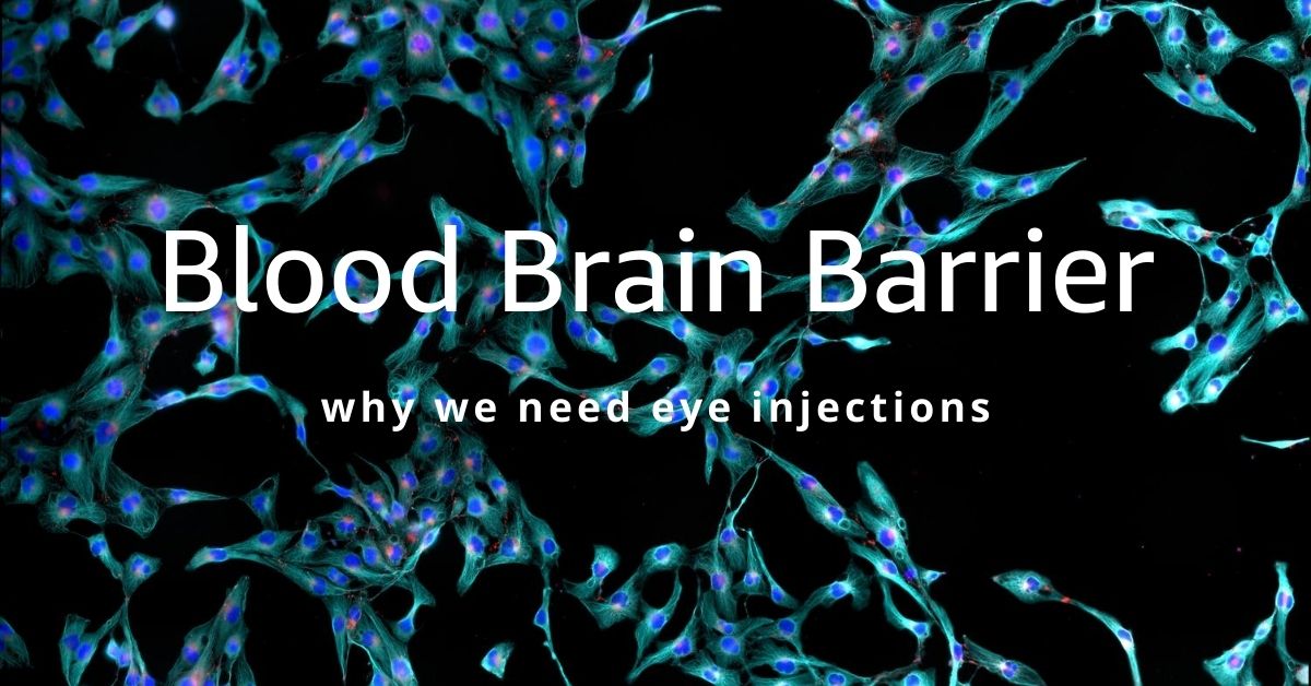
Blood Brain Barrier

The blood-brain barrier (BBB) is a protective layer of endothelial cells that protects the brain from any pathogens or toxins that may be circulating in the blood supply, but it also restricts entry of large-molecule medications and 98% of all small-molecule drugs. So, the only way to get therapeutics to the brain or to the deep structures of the eye is to go through or behind the BBB.

The BBB allows water, some gasses, and fat-soluble molecules to dissolve in the cell membranes and cross its boundary. Alcohol, caffeine, and nicotine easily cross the BBB because they are lipophilic (attracted to fat) and mix easily with the fat in the BBB to gain entry. Oxygen and anesthesia are gasses that can pass through the BBB.

As part of the brain, the BBB prevents therapeutics that have been ingested and are circulating in the blood to gain entry to the eyes. The medication will travel through the body, but almost none of it will reach the eyes.

Leaky Blood Vessels

Wet macular degeneration is caused when blood vessels behind the retina grow abnormally and leak blood and fluid that distorts vision. If left untreated the leaking blood vessels will cause irreversible damage to the photoreceptors that create central vision.

A Shot in the Eye

The only way to get enough medication directly to the retina to stop the growth of the leaky blood vessels is to use an intravitreal injection. An intravitreal injection means that the medication is injected into the vitreous (jelly-like fluid) of the eye where it will diffuse to the retina.

In addition, injecting anti-vascular endothelial growth factor (anti-VEGF) directly into the eye where the leaky vessels are growing, prevents the anti-VEGF from circulating in your blood supply and possibly adversely affecting systemic blood vessels as it moves along to the eye.

The intravitreal injections are painless because the eye is numbed, and the injections can be done in your doctor’s office. They are a safe, and effective way to prevent leaky blood vessel damage to your retina.

Coming Advances in Treatment

An implanted reservoir that delivers sustained-release anti-VEGF directly to the retina is in the experimental phase. And the field of nanotechnology is in the preliminary research stages of developing nanoparticles as a potential way to transfer medication in eye drops across the BBB.

If you would like to schedule an appointment, please call us (877) 245.2020.

Nader Moinfar, M.D., M.P.H.
Retina Specialist
Orlando, FL

Dilated Eye Exams

Why you need to have your eyes dilated by a retina specialist.

In order to properly examine your retina, a dilated eye exam is necessary each and every time you come to the office. A dilated eye exam requires use of dilating drops to dilate and enlarge your pupils to allow us to properly diagnose and treat diseases of the retina.

A dilated eye exam is the only way the retina can be examined by direct visualization using a combination of lenses and specialized ophthalmic instruments.

Other methods such as photography, fluorescein angiography, optical coherence tomography (OCT) and ultrasound can be used in conjunction to direct visualization to diagnose and to treat retinal diseases.

As a retina specialist, virtually everyone of my patients needs a dilated eye exam every time they come to the office.

Dilating Eye Drops

There are several drops used routinely to dilate the eyes. All are marked with a bright red cap to distinguish these eye drops from other types.

In most cases, we only need to use phenylephrine and tropicamide do satisfactorily dilate the pupils. These drops work differently to achieve dilation.

Tropicamide inhibits the small muscle located at the margin of the pupil. The function of the pupillary sphincter muscle is to constrict the iris in bright light and makes the pupil smaller through a purse-string mechanism. Tropicamide prevents this constriction.

Phenylephrine stimulates another set of muscles which function to enlarge the pupil. The iris dilator muscle runs radially along the iris and, when stimulated, pulls the iris open.

Cyclopentolate and atropine can also dilate the pupils but their effects last much longer (days to weeks) and are generally not used for diagnostic purposes.

How Long does Dilation Last?

In most circumstances, your eyes will stay dilated for several hours after the exam. Several factors influence the length of time your eyes will stay dilated, such as:

  • Color of your eyes
  • Your age
  • Type, strength and amount of the medicine
  • Previous eye surgery

Lightly colored eyes, such as blue and hazel, dilate very quickly and stay dilated longer compared to brown eyes.

Brown eyes actually contain brown colored pigment which absorbs the dilating medicine, whereas blue eyes actually contain no pigment and the eyes dilate faster.

Younger eyes tend to take a longer time to dilate. Certain diseases (e.g. iritis) can make dilation take a very long time.

Dilation usually takes at least 20 minutes at a minimum and will reverse after a few hours.

If you would like to schedule an appointment, please call us (877) 245.2020.

Nader Moinfar, M.D., M.P.H.
Retina Specialist
Orlando, FL

Laser Eye Surgery for the Retina

Lasers are commonly used in all of ophthalmology and are useful tools to treat disease of the cornea, glaucoma, cataracts and various retinal diseases.

Because the cornea is transparent to light, the cornea allows the laser energy enter the eye and treat the inner parts of the eye such as the retina.

Lasers for the Cornea

Reshaping the cornea allows many patients to have refractive surgery. Modern refractive surgery is called LASIK. The precision of the laser allows the surgeon to accurately resculp the curvature of the cornea to match the prescription otherwise needed by wearing glasses or contact lenses. An eximer laser is commonly used to perform LASIK.

Laser for Glaucoma

Laser surgery for glaucoma has been long been a treatment option to reduce intraocular pressure. Reducing the internal eye pressure is the mainstay of most treatments for glaucoma. Argon, diode or Neodymium:YAG lasers can be used for glaucoma laser surgery.

Cataract Laser Surgery

There are two ways lasers can assist the eye surgeon to treat cataracts and so-called after cataracts. Laser assisted cataract surgery allows the eye surgeon to more accurately perform several steps of the cataract surgery, but not the entire surgery. A femto-second laser is used for laser-assisted cataract surgery.

After initial cataract surgery, the intraocular lens commonly becomes cloudy. This cloudiness can be improved with a Nd:Yag laser.

Retina Laser Eye Surgery

Lasers are used by a retina specialist to treat 3 different types of problems:

A retinal tear can happen to anyone. Retinal tears can cause a retinal detachment which requires surgery and is vision threatening. Using an argon or diode laser, laser burns surround the retinal tear to prevent a retinal detachment from occurring from that specific tear. In essence, the retinal burn, “seals” the tear, preventing retinal detachment.

Certain types of retinal vascular occlusions (RVO) can cause swelling, aka edema, in the retina. Similar types of swelling are commonly treated in patients with diabetes. The laser acts to decrease swelling by either reducing the rate of fluid accumulation or increasing the rate of absorption.

Advanced retinal diseases, called retinopathy, can be treated with laser to stabilize the eye and prevent vision loss. Proliferative diabetic retinopathy is often treated with laser or intravitreal injections. Retinopathy can also be caused by types of vascular occlusions.

If you would like to schedule an appointment, please call us (877) 245.2020.

Nader Moinfar, M.D., M.P.H.
Retina Specialist
Orlando, FL

Jon Doe