There is a variety of surgery to repair a retinal detachment.
Retinal detachments are potentially blinding and usually require surgery. Retina specialists are the type of eye doctor who specialize in this type of surgery.
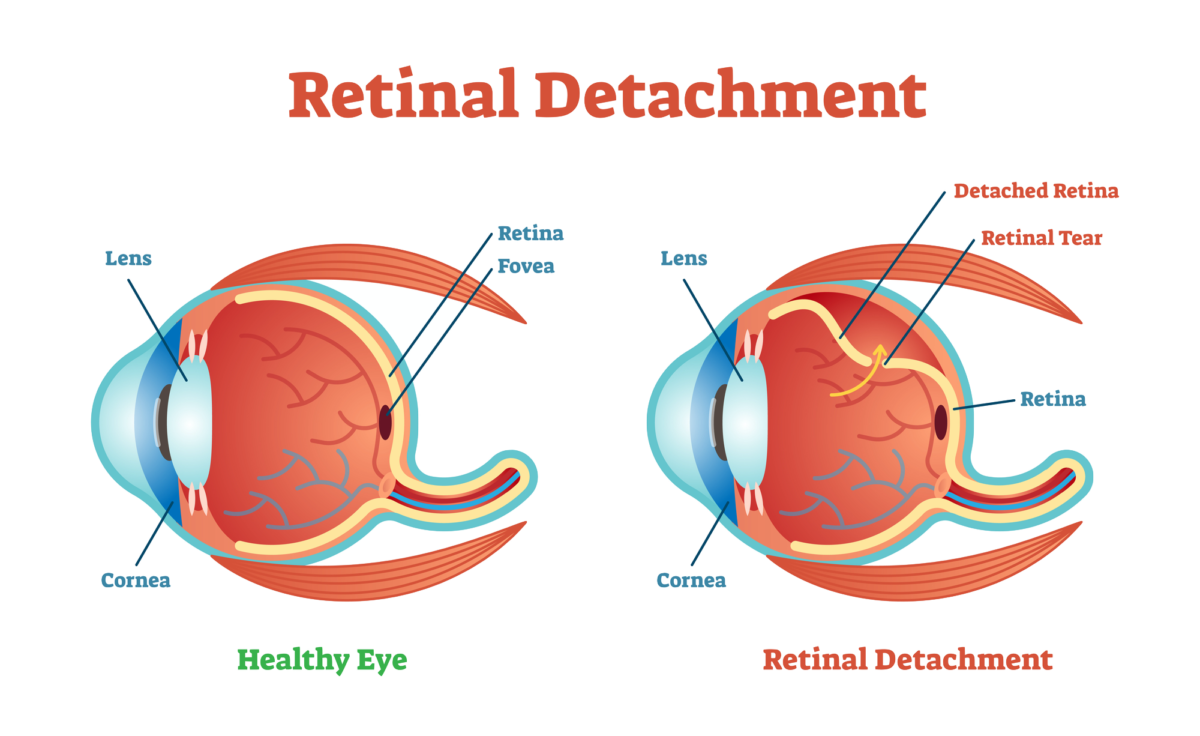
The retina is the electrical circuitry of our eyes, and it covers about 65% of the back of the eye. It contains rod and cone photoreceptor cells that are biological transducers, which means the cells translate physical stimuli (the light reflected off objects) into electro-chemical signals that are sent to the visual cortex in the brain via the optic nerve where they create our vision.
When the retina has a tear or a partial detachment that section of the circuitry can no longer send visual signals. There is no visual input from the torn or detached area.
Retinal detachment surgery may be a medical emergency and must be treated quickly. The longer and greater the extent of the retinal detachment, the worse the visual outcome.
Symptoms of Retinal Detachment
- Flashes of light
- Lots of new floaters (small black specks or threads) in your vision.
- A shadow or “curtain” descending from the top or across the side of your eye
Some retinal detachments happen gradually and are preceded by flashes of light and floaters and others detachments happen rapidly and the only symptom is the descending dark curtain blocking vision where the retina has detached.
Causes
An injury to the face or eye can detach a retina. Aging can cause changes in the gel-like vitreous material inside the eye. When the changed vitreous moves around it can tug on the retina and cause a detachment. Tumors and some diseases such as diabetes can cause retinal detachments.
A posterior vitreous detachment (PVD) is a common cause of retinal tears leading to retinal detachment.
Treatments
Pneumatic Retinopexy is the simplest fix for small retinal detachments and the procedure can be done in the office. With your eye numbed, a gas bubble is injected into your eye so that it presses against the detachment to hold it in place and then the area is sealed using a cryogenic (freezing) probe or a laser. After the procedure you must keep your head in the position recommended by your doctor for about 1 to 3 weeks.
Scleral buckling is a surgical procedure performed in the operating room. The sclera is the white outer layer of the eye and your surgeon attaches a band made of silicone around the sclera from top to bottom. This compresses the eye and pushes the retina back into place where it will reattach to its blood supply and heal.
Vitrectomy is the removal of the vitreous gel that fills the eye. The retina specialist removes any scar tissue and seals any retinal tears and then fills the eye with saline, air, or a gas bubble. Over time your eye displaces and replaces them with its own fluid. The vitreous does not regenerate, but your eye is able to function well without it.
Retina specialists vary in the their approach to retinal detachment surgery, and often, a combination of the methods and techniques that are described above can be utilized.
Recovery
Vision will gradually improve after retina reattachment. It may take up to six months or longer to regain your best vision. An air or gas bubble injected into the eye will temporarily blur your vision until the bubble dissipates.
Less vision will be recoverable If the detachment was severe and the central portion of the retina was detached, or if treatment was delayed and the retina was without blood flow for an extended time.
Not all retinal detachments situations are the same and it is recommended you communicate with your own doctor about your prognosis.
If you would like to schedule an appointment, please call us (877) 245.2020.