7 Reasons You Need a Retina Specialist

A retina specialist has undergone expert training to deal with diseases and surgery of the retina and vitreous such as these disorders or situations.

A retina specialist is an ophthalmologist who has extra-training in diseases and surgery of the retina. Additional training years are required to focus attention in this special area.

Here are 7 more common reasons why you may be referred to a retina specialist.

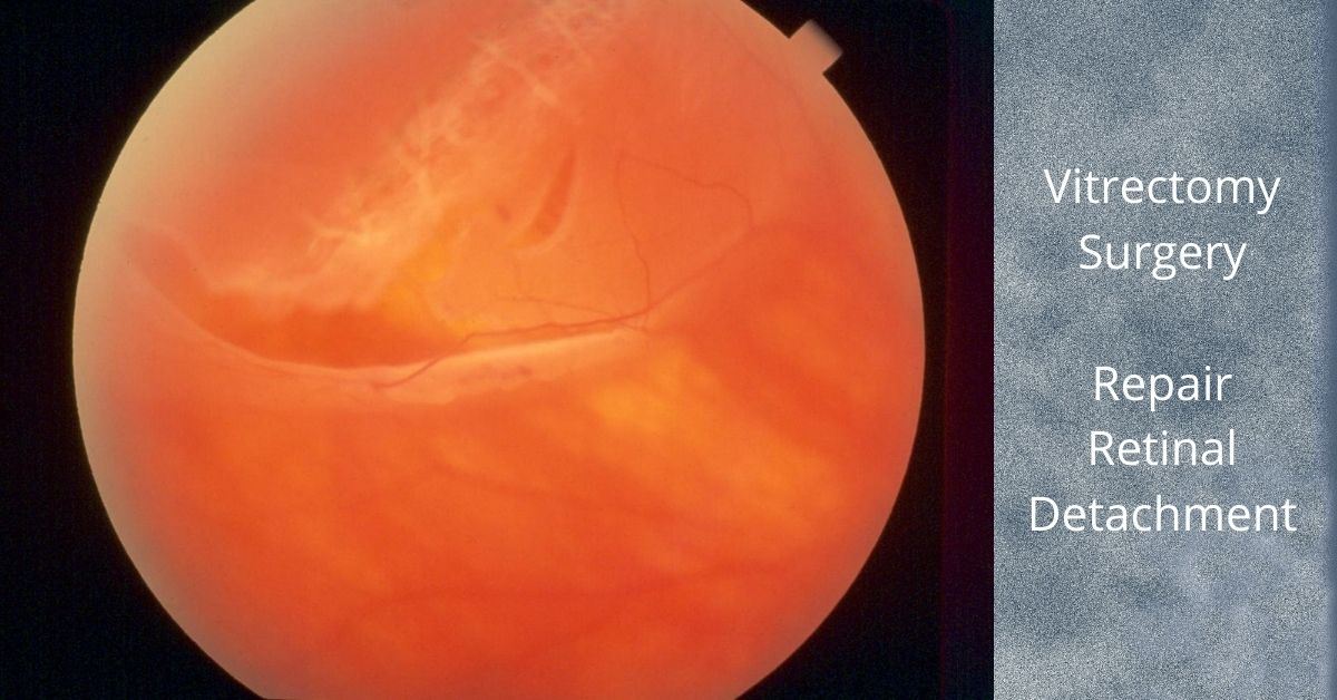
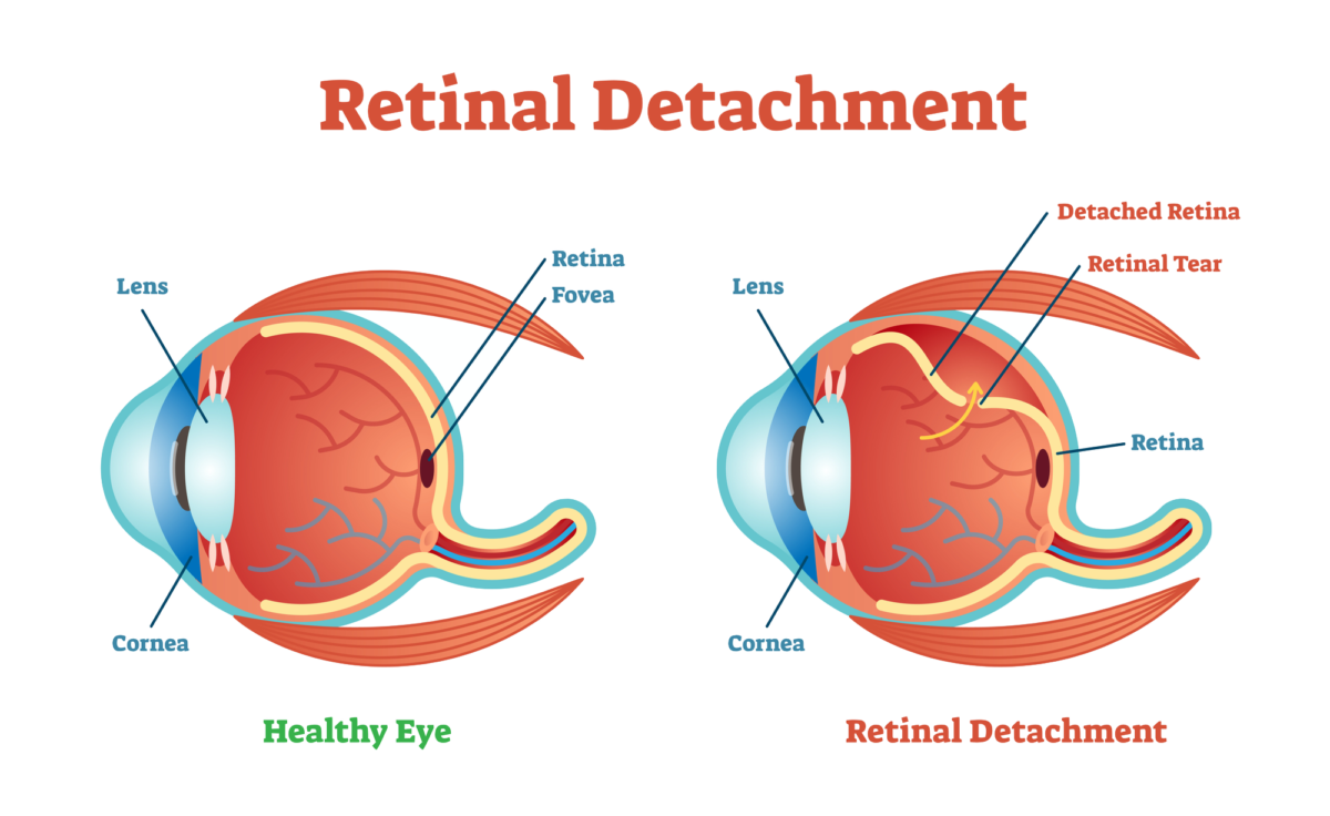
Retinal Tear, Retinal Detachment, Retina Specialist

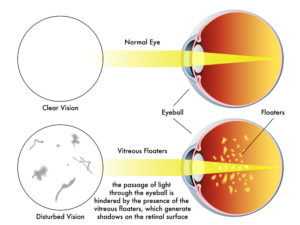
Retinal tears can cause a retinal detachment. Retinal tears may be asymptomatic, but often are associated with the sudden onset of new floaters or flashes usually due to a posterior vitreous detachment.

While most new floaters and flashes are NOT associated with a retinal tear, the only way to make sure you are not at risk for developing a retinal detachment is have a complete dilated eye exam.

Retinal tears can be treated with either cryotherapy or laser. Retinal detachments will often require surgery.

Foreign Body Inside the Eye

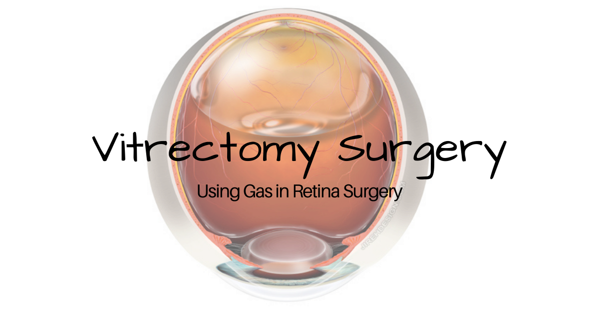
While not commonplace, intraocular foreign bodies may occur with any penetrating eye injury. Glass, metals and organic material need to be removed by performing a vitrectomy, the principal operation performed by a retina specialist.

Retina Specialist and Endophthalmitis

Endophthalmitis is infection inside the eye. This usually occurs following recent eye surgery, but can also follow penetrating eye injury.

Prompt referral to a retina specialist is paramount in preventing loss of vision from infection. Intraocular antibiotics, anti-fungals and/or vitrectomy surgery may be necessary to prevent catastrophic vision loss.

Advanced Diabetic Retinopathy

Diabetic retinopathy can cause blurry vision and blindness. A retina specialist is adept at treating the two most common causes of vision loss from diabetes: diabetic macular edema and proliferative diabetic retinopathy.

Common treatments used by retina specialists include: anti-VEFG injections, steroids, laser and surgery. In most scenarios, these are procedures only offered by a retina specialist.

Macular Degeneration

Macular degeneration (AMD) comes in two forms: dry AMD and wet AMD. The “wet” form is also called neovascular AMD (nAMD).

Both types of macular degeneration affect both eyes and are progressive, but the wet form can steal vision quickly, is associated with leaky abnormal blood vessels and bleeding. Treatment usually involves intravitreal injections of anit-VEGF.

There is presently no treatment for dry macular degeneration approved by the FDA.

Retinal Vascular Occlusions

Periodically the retinal vessels can become occluded or blocked. Both arteries and veins can be blocked retinal vascular occlusions. Vision loss is usually sudden and treatments may be required in an attempt to improve vision and/or to prevent total blindness from complications of the vascular occlusions.

If you would like to schedule an appointment, please call us (877) 245.2020.

Nader Moinfar, M.D., M.P.H.
Retina Specialist
Orlando, FL

Vitrectomy for Retinal Detachment Repair

This is the most common way to repair most retinal detachments and has faster healing times.

Vitrectomy for retinal detachment repair is an alternative method to scleral buckle or pneumatic retinopexy.

A vitrectomy is a common surgical repair for a detached retina. The surgery removes the vitreous gel from the eye, pushes the detached retina back into place, and seals any retinal tears in the retina. Then gas is injected into the eye to help keep the retina in place and prevent any fluid from leaking from the repaired tears and into the subretinal space.

The Surgery

A vitrectomy is usually performed using local anesthetic. A heavy sedative is administered and then Lidocaine is injected around the eye to numb it. If patients prefer not to be awake during the procedure they can be given intravenous sedation or a general anesthesia.

The surgery consists of making three small needle-sized incisions (about the width of an eyelash) into the white part of the eye (sclera). Through one incision a constant flow of fluid is passed into the eye. The second incision is used to insert a fiber-optic light to provide illumination of the eye, and the third is for any instruments that are used, including a “vitrector” for the removal of the vitreous.

A vitrector is an oscillating microscopic cutter that slowly removes the vitreous. Once the vitreous is removed, a freezing instrument or a laser is used to seal any tears in the retina.

The vitreous will be replaced with a gas. In time the gas will dissipate and be replaced by the eye’s own fluids (aqueous humor). The vitreous gel does not grow back, but the eye can function with just the fluid that replaces the vitreous. Depending on the type of gas used and its concentration, it can take from three to ten weeks to fully dissipate.

Most retina specialists will perform vitrectomy for retinal detachment repair in an outpatient setting. The surgery time ranges from 20 to 40 minutes, but varies greatly.

Head positioning

Head positioning after surgery is necessary to keep the gas bubble in the proper place. If the retina detached at the bottom, the only way to get the bubble to press against the place of detachment is to place the head down. The bubble walls off the damaged area while it heals and prevents any fluid from flowing through the damaged area of the retina and into the subretinal space.

The gas bubble wiur vision while it is in place, but as it dissipates a line will form across your vision where the newly forming fluid (aqueous humor) is gradually replacing the bubble. The line will move lower each day and the field of vision will get larger.

If you have to position your head down there are pillows, chairs, and mirrors that allow you to see around the room while in a face-down position.

Gas bubble precautions

As long as the gas bubble remains in your eye, you must not fly in an aircraft. The reduced pressure in the cabin will cause the gas bubble to expand. This would be extremely painful and could lead to loss of sight. If you must have general anesthesia for any surgical procedure while the gas bubble is in your eye, you must inform your anesthesiologist so you will not be given nitrous oxide, which would cause a dangerous rise in eye pressure in the eye with the gas bubble.

Safety

Vitrectomy surgery was first developed in 1970 and since then many improvements in instrumentation and technique have dramatically improved the safety of the procedure. Severe complications are rare and the surgical success rate is over 90%.  

Nader Moinfar, M.D., M.P.H.
Retina Specialist
Orlando, FL

If you would like to schedule an appointment, please call us (877) 245.2020.

What is Scleral Buckle Eye Surgery

Scleral buckle eye surgery has been around for generations and is still a mainstay of retinal detachment surgery.

Scleral buckle eye surgery to repair a retinal detachment has been in use since the early 1950s and it remains an essential technique for reattaching the retina.

The scleral buckle, which is usually made of silicone semi-hard plastic, supports the retina from outside the eye. It is placed under the eye muscles and usually encircles the sclera or white part of the eye and eliminates the tugging that is detaching the retina. 

The buckle is placed behind the eyelids and cannot be seen. It is sewn to the sclera (the actual white part of the eye) and it presses the walls of the eye inward, taking pressure off the retina and allowing it to reattach to the eye’s interior wall. The buckle usually remains in place permanently.

Scleral buckling is sometimes a stand-alone treatment for the repair of a retinal detachment and sometimes it is used in combination with a vitrectomy.

A laser or a sub-zero probe (cryopexy) may also be used to seal off the areas of the retina that were torn to prevent further fluid leakage and pressure on the retina.

Side effects of a scleral buckle

The possible complications or side effects of scleral buckling surgery include:

  • Increased myopia (nearsightedness) caused by the increased length of the eye when it is buckled
  • Increase in astigmatism also caused by the buckle changing the eye’s shape
  • Double vision is uncommon but can occur because the eye’s muscles were manipulated when placing the buckle under the eye muscles.
  • Pain after the procedure which can usually be controlled with acetaminophen or ibuprofen and will subside in a few days.

 What causes retinal detachment?

The most common type of retinal detachment, that accounts for 90% of all retinal detachments,occurs when a small tear or hole appears in the retina that allows the vitreous gel of the eye to seep behind the retina where it pushed the retina away from the back of the eye. 

Tears in the retina are caused by vitreous gel, which contains millions of fibers, that becomes “sticky” and tugs at the retina making minute tears in it. Retinal holes develop in areas where the retina has thinned. Retinal holes are typically smaller than tears and have a much lower risk for causing a retinal detachment.

Recovery after scleral buckling eye surgery

The recovery time varies, but ranges from two-to-eight weeks. An eye patch may be required for a few days after surgery and antibiotic eye drops may be prescribed to prevent infection. Your eye may have redness, tenderness, and swelling for a few weeks after surgery.

If you would like to schedule an appointment, please call us (877) 245.2020.

Nader Moinfar, M.D., M.P.H.
Retina Specialist
Orlando, FL

What Causes a Macular Hole?

Retinal surgery sometimes uses injection of gas into the eye, especially when you have a macular hole.

Causes of a macular hole are discussed in this article and explained in the embedded video.

A macular hole is a hole at the very center of the retina. The retina is the layer of tissue that lines the inside of the eye and it contains millions of light-sensitive cells that receive and send visual information to the brain.

The macula portion of the retina contains the highest concentration of light-sensitive cells and is responsible for high-resolution, detailed central vision and most of our color vision.

Holes in the macula can be caused by injury, but most macular holes occur in people over the age of 60 and are caused by the vitreous gel in the eye pulling on the macula. These are called idiopathic macular holes and are, for reasons unknown, more common in women than men.

The Role of the Vitreous in Macular Holes

The vitreous contains millions of microscopic fibers that attach to the retina. As people age the vitreous slowly shrinks and pulls away from the retina’s surface and natural fluid fills in the area where the vitreous contracted. This is normal and usually causes no problems beyond possibly seeing “floaters” in your visual field from time to time.

However, in some cases the vitreous is so firmly attached to the retina that when it pulls away it can tear the retina slightly and cause a hole to form. Small holes sometimes heal on their own, but they can also gradually increase in size causing vision loss.

Other Causes of Macular Hole

The following additional conditions can cause a macular hole to develop:

  • Blunt trauma to the eye
  • Diabetic eye disease 
  • High degree of myopia (nearsightedness)
  • Macular pucker—caused by scar tissue on the macula

Symptoms of a Macular Hole

In the early stages there may be a slight distortion or blurriness in central vision. As the hole increases in size, straight lines and objects look bent or wavy, vision becomes increasingly blurrier and a dark spot may appear in the center of your vision.

Treatment of Macular Holes

A surgery to remove the vitreous gel (vitrectomy) and prevent it from continuing to pull on the retina is currently the best way to repair a macular hole. After the removal of the vitreous gel, a bubble containing a mixture of air and gas is put into the eye to prevent subretinal fluid from seeping behind your retina and destabilizing the healing process.

The gas bubble will slowly dissipate and be replaced with aqueous humor produced by your eye. You may be asked to keep your head in a face-down position for several days to keep the bubble in place. CAUTION: As long as any of the gas bubble remains in your eye you must not fly in an airplane because the bubble can expand in the reduced pressure of the cabin causing severe pain and possible loss of sight.

Another potential treatment for some patients with macular holes is the injection of an antiplasmin inhibitor that inactivates plasmin, an enzyme that breaks down the fibrin in blood clots.

Success Rate

The vitrectomy success rate is over 90% with patients regaining most of their lost vision. The gas bubble starts to shrink 7 to 10 days after the vitrectomy, but it takes about 6 to 8 weeks for the gas bubble to be totally absorbed. Vision will continue to improve during that 6 to 8 week time.

Complications

In less than 10% of cases the vitrectomy may cause cataract formation, retinal detachment, infection, glaucoma, bleeding or a re-opening of the macular hole. 

Nader Moinfar, M.D., M.P.H.
Retina Specialist
Orlando, FL

If you would like to schedule an appointment, please call us (877) 245.2020.

Gas Bubbles and Retina Surgery

Not all retina surgery requires a gas bubble, but here’s when it is necessary for your retina specialist to use a gas.

Not all retina surgery requires injection of a gas bubble.

To safely work on the retina, your retina specialist must be able to safely access the inner layer of the eye. A vitrectomy, the procedure to remove the vitreous, allows the retina specialist specialist to work on the retina. 

A vitrectomy (removal of the vitreous humor gel) is done for the following conditions:

The Gas Bubble

A tamponade in medical terms is something used to close or block a wound or body cavity to stop bleeding or fluid leakage.

Not all retina surgery requires gas to be injected into the eye. The most common use of gas is for repair of a retinal detachment or macular hole. In the case of the retinal detachment (specifically a rhegmatogenous retinal detachment which is caused by a retinal tear or retinal hole), the gas is used to block (tamponade) the migration of fluid to through the tear to underneath the retina.

In the case of a macular hole, the gas is used to allow the hole to slide closed by surface tension.

The gases commonly used are sulfur hexafluoride (SF6) and perfluoropropane (C3F8), and air. Each of the gases dissipates and is replaced by natural fluid, but the time for complete dissipation varies. Air dissipates and is replaced by natural fluid in 5 to 7 days. Sulfur hexafluoride (SF6) dissipates in 10 to 14 days, and perfluoropropane (C3F8), in 55 to 65 days.

The gas bubble blurs your vision while it is in place. As the bubble dissipates you will see a line across your vision where the gas meets the newly forming fluid which is gradually replacing the bubble. The line will move lower each day and your field of vision will get larger as the natural fluid continues to replace the bubble.

As long as any of the gas bubble remains in your eye you must not fly in an airplane because the bubble can expand in the reduced pressure of the cabin causing severe pain and possible loss of sight.

Head Position

Your surgeon will ask you to position yourself in a specific way during healing and that position is dependent on what part of the retina was repaired.

In cases of macular holes, a face down position is common. Head positioning for retinal detachments depends upon the location of the retinal tear(s).

Gas Bubble Injected Last

Most retina surgery is outpatient surgery and can be done under local anesthetic and mild sedation. The sedation is given by IV and is used for anxiety relief and to put you into a relaxed and sleepy state, known as a “twilight state”. In that state you are conscious and still able to hear and follow simple instructions from your surgeon.

General anesthesia can be used for patients with dementia, severe anxiety, or young children.

Most retina surgeries take less than an hour and some less than 30 minutes. The gas bubble is injected as one of the last steps of the surgery.

Recovery

You will have to wear an eye patch for a day or two following surgery. Recovery time depends on the procedure you had, but is generally two to four weeks. An exception is the repair of a complete retinal detachment which could take several months to heal and for vision to stabilize.

If you would like to schedule an appointment, please call us (877) 245.2020.

Nader Moinfar, M.D., M.P.H.
Retina Specialist
Orlando, FL

Surgery for Retinal Detachment

Here’s how a retinal detachment occurs and the various surgical methods used by a retina specialist to repair this potentially blinding problem.

There is a variety of surgery to repair a retinal detachment.

Retinal detachments are potentially blinding and usually require surgery. Retina specialists are the type of eye doctor who specialize in this type of surgery.

The retina is the electrical circuitry of our eyes, and it covers about 65% of the back of the eye. It contains rod and cone photoreceptor cells that are biological transducers, which means the cells translate physical stimuli (the light reflected off objects) into electro-chemical signals that are sent to the visual cortex in the brain via the optic nerve where they create our vision.

When the retina has a tear or a partial detachment that section of the circuitry can no longer send visual signals. There is no visual input from the torn or detached area.

Retinal detachment surgery may be a medical emergency and must be treated quickly. The longer and greater the extent of the retinal detachment, the worse the visual outcome.

Symptoms of Retinal Detachment

  • Flashes of light
  • Lots of new floaters (small black specks or threads) in your vision.
  • A shadow or “curtain” descending from the top or across the side of your eye

Some retinal detachments happen gradually and are preceded by flashes of light and floaters and others detachments happen rapidly and the only symptom is the descending dark curtain blocking vision where the retina has detached.

Causes

An injury to the face or eye can detach a retina. Aging can cause changes in the gel-like vitreous material inside the eye. When the changed vitreous moves around it can tug on the retina and cause a detachment. Tumors and some diseases such as diabetes can cause retinal detachments.

A posterior vitreous detachment (PVD) is a common cause of retinal tears leading to retinal detachment.

Treatments

Pneumatic Retinopexy is the simplest fix for small retinal detachments and the procedure can be done in the office. With your eye numbed, a gas bubble is injected into your eye so that it presses against the detachment to hold it in place and then the area is sealed using a cryogenic (freezing) probe or a laser. After the procedure you must keep your head in the position recommended by your doctor for about 1 to 3 weeks.

Scleral buckling is a surgical procedure performed in the operating room.  The sclera is the white outer layer of the eye and your surgeon attaches a band made of silicone around the sclera from top to bottom. This compresses the eye and pushes the retina back into place where it will reattach to its blood supply and heal.

Vitrectomy is the removal of the vitreous gel that fills the eye. The retina specialist removes any scar tissue and seals any retinal tears and then fills the eye with saline, air, or a gas bubble. Over time your eye displaces and replaces them with its own fluid. The vitreous does not regenerate, but your eye is able to function well without it.

Retina specialists vary in the their approach to retinal detachment surgery, and often, a combination of the methods and techniques that are described above can be utilized.

Recovery

Vision will gradually improve after retina reattachment. It may take up to six months or longer to regain your best vision. An air or gas bubble injected into the eye will temporarily blur your vision until the bubble dissipates.

Less vision will be recoverable If the detachment was severe and the central portion of the retina was detached, or if treatment was delayed and the retina was without blood flow for an extended time.

Not all retinal detachments situations are the same and it is recommended you communicate with your own doctor about your prognosis.

If you would like to schedule an appointment, please call us (877) 245.2020.

Nader Moinfar, M.D., M.P.H.
Retina Specialist
Orlando, FL

Symptoms of a PVD

These symptoms may prevent a retinal detachment and here’s why.

PVD stands for “posterior vitreous detachment.” It’s a normal event that increases your risk for developing a retinal tear which can lead to a retinal detachment. Everyone will eventually get a PVD.

New flashes and new floaters should be reported to your eye doctor or retina specialist. Not all floaters are benign. Only a full dilated exam will determine if your new symptoms are related to a posterior vitreous detachment, retinal tear or something else.

Symptoms of Posterior Vitreous Detachment

The most common symptoms of a PVD:

  • Flashes
  • Floaters
  • Nothing (Asymptomatic, no flashes or floaters)

There are two ways to stimulate the retina: normal light and physical manipulation. Usually, light enters the eye and is captured by the rods and cones of the retina which eventually produce vision. The other mechanism occurs when you rub your eyes so hard you actually see patterns in your vision. This rubbing, or physical manipulation, also produces “light.”

After a PVD has occurred, only the back, or posterior portion, of the vitreous separates from the retina. The anterior portion remains fixed to the surface of the retina. As the newly separated vitreous cleaves from the retinal surface in the back, it can move to and fro in the eye. This transmits energy to the portion of the vitreous still attached to the retina in the anterior part of the eye…causing flashes.

What are Floaters?

Floaters can be a result of cellular debris from underneath the retina in cases of a retinal tear, blood or just opacities which have formed in the otherwise clear vitreous gel.

Not all posterior vitreous detachments cause flashes or floaters. Thus, follow up is always recommended so your retina specialist can look for asymptomatic retinal tears.

What if I have a Retinal Tear?

First of all new flashes and floaters should be reported to your eye doctor. In the case of a PVD, it’s important to look for a retinal tear. A retinal tear is most likely to occur during the first 6 weeks after a PVD has occurred.

The goal of examination is to find a retinal tear before it turns into a retinal detachment. Retinal tears can usually be treated with laser whereas a retinal detachment requires surgery and there is a chance of lost vision.

If you would like to schedule an appointment, please call us (877) 245.2020.

Nader Moinfar, M.D., M.P.H.
Retina Specialist
Orlando, FL

Flashes and Floaters

Here’s what you need to know about those pesky floaters and why they could blind you.

Eye “floaters” are deposits or debris within your eye’s vitreous humor. Whether you see specks, wispy threads, strings, or cobweb shapes that drift back and forth when you move your eyes, you are seeing floaters. More precisely, you are seeing the shadows that the floaters cast on your retinas. Floaters can refract light and that temporarily interferes with your vision until the floaters drift into a different position.  

Harmless Floaters?

Eye floaters move around, come and go, and eventually settle away from the line of sight after a few weeks. They can be annoying and distracting, but most of the time they are harmless and are not a cause for alarm, however, any sudden onset of new floaters should be examined.

Floaters are suspended in the vitreous humor which fills about 80 percent of the eye. The most common cause of floaters is shrinkage of the vitreous humor. The vitreous humor is 98 to 99% water and the remaining 1 to 2% is a mixture of collagen, proteins, salts, and sugars. Despite the water-to-collagen ratio, the vitreous humor has a firm jelly-like consistency.

Posterior Vitreous Detachment

As we age the vitreous humor shrinks and liquefies and develops a watery center. Portions of the collagen and protein mixture become stringy fibers and these floating stringy collagen and protein fibers are the floaters. This is called a PVD (posterior vitreous detachment).

Sometimes the small floating vitreous fibers pull on retinal cells and that mechanically stimulates the retina cells causing the sensation of flashes of light. This too is not unusual. Floaters and flashes occur at the same time and are mostly harmless annoyances. You have experienced mechanical stimulation of retinal cells if you’ve ever been hit in the eye and afterwards “see stars”.  

Treatments

In rare instances floaters can be so closely packed and large or so numerous that they significantly affect vision. In those cases, floaters can be treated with a surgical vitrectomy or a laser vitrectomy.  A surgical vitrectomy removes the vitreous humor and replaces it with saline or a bubble made of gas or silicone oil. A laser vitrectomy breaks apart large floaters and vaporizes them.

Floaters and flashes of light that require immediate attention

A sudden onset of numerous floaters, frequent flashes of light, a dark shadow that covers part of your side vision, or eyes that hurt can be symptoms of a tear in your retina and could indicate that your retina is detaching. A detached retina is an emergency and immediate treatment can save your sight. 

If you want to make an appointment, please call us (877) 245.2020.

Nader Moinfar, M.D., M.P.H.
Retina Specialist
Orlando, FL

Treatment of Floaters

“Floaters” is a very broad term, describing objects that seem to float in your vision. By far and away, floaters are not dangerous nor sight-threatening– they are simply changes in the jelly of the eye, called the vitreous, that occurs as we grow older.
In some instances, floaters can represent blood or pigment, that may arise from a retinal tear; you can read more about this under the “retinal tears” section of NaderMoinfarMD.com.
Assuming that the floaters are not from a retinal tear, inflammation or certain cancers of the eye, it is perfectly fine to just leave floaters alone. Over time, much of the visual disturbance will simply diminish, as the floater breaks up and becomes smaller.
I personally have had floaters in both eyes since college, and really don’t even notice them very much. Like most people, I will notice them if I’m reading, or observing something against a white background.
Occasionally, a patient may be referred to me for floaters that are truly interfering with their activities of daily living– pilots, professional drivers, etc. Typically, persons such as these will have a large opacity that is pretty much stuck in their central vision, causing a blind spot– perhaps posing a danger to themselves, and well as others.
Treatment for floaters is widely discussed, and there are really no great clinical studies or randomized trials to suggest what works best. You may read some who advocate doing laser, and some who even offer nutritional advice.
I would suggest that if you are concerned, that you seek the help of a fellowship-trained retina specialist. An experienced retina surgeon can offer the most definite treatment, which is to physically remove the floaters through a procedure called a vitrectomy. You can view videos of how a vitrectomy is performed under the “videos” section of NaderMoinfarMD.com.
Briefly, a vitrectomy is an outpatient procedure performed under local anesthesia. The vitreous is removed, and temporarily replaced with saline; over time, your eye will replace the saline with its own fluid. Recovery time is just a day or two, and patients can usually resume most of their regular activities soon thereafter. If performed correctly, once the floaters/junk are removed, they should not come back.

Treatment of a Retinal Detachment

Retinal Detachment | Nader Moinfar MD | Lake Mary Orlando| Retina Specialist
A total retinal detachment.

A retinal detachment usually when there is pulling on the retina, which causes a tear to occur. Causes of retinal tears are multiple, and include trauma, genetic predisposition, myopia (near-sightedness), to name a few.

A retinal tear should be evaluated by a trained retina specialist, and can usually, and successfully, be simply treated in the office.

A retinal detachment is a more complicated problem, and a well- trained retina specialist can offer you different ways of treating this potentially blinding condition.

In some cases, an office-based procedure called a pneumatic retinopexy can be offered; with appropriate selection of the patient, success rates of up to 80% can be achieved. In the office, a gas bubble is injected into the eye; and either the same day, or maybe a few days laser, laser or cryotherapy is applied. Your retina specialist should discuss with you his or her experience level with this procedure, success rates, and experience levels with the other options, that may be available.

Another method to treat retinal detachments, as seen under the video section of NaderMoinfarMD.com, is a procedure called a vitrectomy. Generally speaking, the vitreous, which is the clear jelly of the eye, is the cause of a retinal tear and traction. By removing the vitreous, one removes the underlying cause. A vitrectomy for a retinal detachment must be careful and completely done, to ensure the greatest chance of success. A seen in my videos, the vitreous is removed, and following a procedure termed an air-fluid exchange, laser is applied around the tears, and finally, the space is filled with a gas bubble.

Lastly, your retina specialist may advise alone, or in combination with a vitrectomy, a procedure termed a scleral buckle; this, too, can be viewed on my video section. With this procedure, a very thin band of silicone is secured on the outside, white part of the eye, helping to relieve the internal traction on the retina; cryotherapy, and a gas bubble, are also used.

With almost all cases of retinal detachments, you will be required to keep your head and body in a certain configuration for a short time; this allows the gas inside your eye to push up against the detachment, allowing it to heal, or “stick’ down. Please note that when the gas bubble is in the eye, you should not travel by air, go up to certain altitudes, and must avoid certain types of general anesthetic inhalation agents if having surgery. Your retina specialist should provide you with a green wrist band to remind you, and others, of this precaution.

If you want to make an appointment, please call us (877) 245.2020.

Nader Moinfar, M.D.
Retina Specialist
Orlando, FL

Jon Doe