Drusen are small deposits of lipid, a type of fatty material, found within the layers of the retina. They are often associated with macular degeneration, but they are not sine qua non of making the disease.
Drusen are not necessarily indicative of macular degeneration (AMD). Both wet and dry forms of AMD can have these tiny white dots in the retina.
Types of Drusen
Drusen can also be found in the optic nerve, but they are different than those found in the retina.
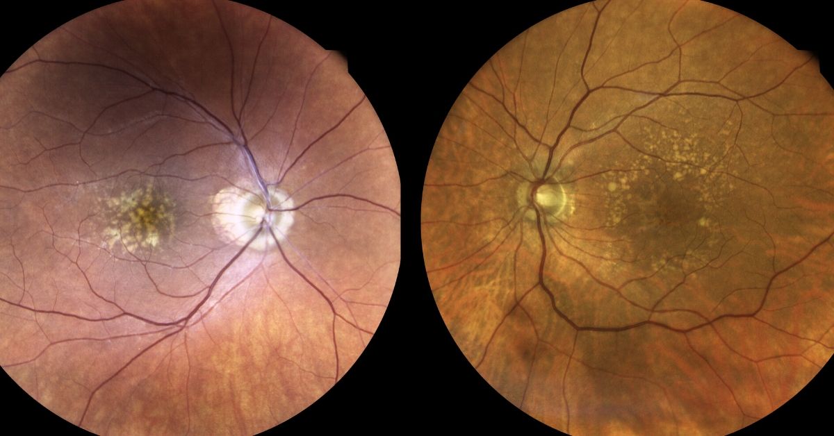
Drusen of the retina are classified as either soft or hard. The distinction is based upon their appearance only. Hard druse are smaller, increase with age and are less often associated with macular degeneration. Both hard and soft are yellow/white in color.
Soft druse tend to be found in clumps or even run together. There is a stronger association between soft drusen and the development of AMD.
Drusen, when not found in the macula, are usually of little consequence and are not considered in the diagnosis of macular degeneration.
Diagnosis of Macular Degeneration
There are several criteria contributing to the diagnosis of macular degeneration:
- Age – usually over 50-55 years old
- Race – AMD is much more common in patients of northern European descent.
- Appearance – or signs of the disease include pigment changes of the retina, fluid, leakage, blood, scarring, +/- drusen
- Symptoms – all patients with macular degeneration have decreased vision and/or distortion. AMD usually involves both eyes.
The point is that drusen may hint of macular degeneration but they alone do not define (make the diagnosis) AMD. There must be additional findings and symptoms.
Testing for Macular Degeneration
If your eye doctor is suspicious of macular degeneration, he/she may recommend periodic examination and home monitoring. Home monitoring may consist of using an Amsler grid.
Referral to a retina specialist may also be considered.
Additional testing, often performed by a retina specialist, may include fluorecein angiography, photography or optical coherence tomography (OCT).
If you would like to schedule an appointment, please call us (877) 245.2020.