Syfovre™ for Geographic Atrophy

Syfovre™ is the first available FDA approved treatment for geographic atrophy, an advanced form of macular degeneration.

Syfovre™ is a novel treatment for an advanced stage macular degeneration called geographic atrophy.

The FDA has approved Syfovre™ (pegcetocplan) for the treatment of GA, a leading cause of in developed countries. Syfovre™ is the first treatment for this advanced stage of macular degeneration. Treatment for wet macular degeneration has been available for over a decade.

Administration of Syfovre™

As with many retinal therpies, Syfovre™ is administered as an intravitreal injection into the eye. Because eye drops, ointments, oral meds and intravenous medications have difficulty penetrating the eye, intravitreal injections are necessary to deliver the right amount of drug to the retina.

Injections may be given monthly or every other month and will need to be repeated. The exact frequency of dosing will be determined by your doctor.

How Does Syfovre™ Work?

Pegcetocoplan is a unique inhibitor of C3, a protein involved in the complement system. The complement system is a major factor in our immune system and helps prevent infection from bacteria, viruses, regulates inflammation and aids the body get rid other pathogens.

Left unchecked, C3 may be involved in the death of healthy cells of the retina called retinal pigment epithelial (RPE) cells. RPE cells are responsible for the health and maintenance of photoreceptors, such as rods and cones. Without healthy photoreceptors, we can not see.

Syfovre™ binds C3 much like an antibody and prevents C3 from causing cell death to the retinal RPE cells.
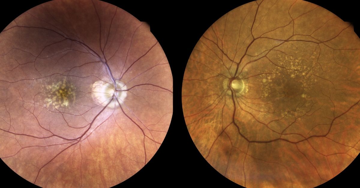
Geographic Atrophy

Areas of RPE cell loss can be detected during a retinal examination. Usually these so-called “atrophic lesions” arise near the macula. With time the lesions enlarge to involve the central macula.

Visual acuity, your central vision, is usually spared early in the disease, but visual function may be compromised depending upon the size, number and location with respect to the central macula.

Unlike wet macular degeneration where abnormal blood vessels develop within the layers of the retina, geographic atrophy is causes by death of the RPE cells.

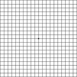
Symptoms of Geographic Atrophy

Symptoms of macular degeneration include: blurry vision, distortion and loss of central vision. Geographic atrophy can also cause decline in visual function such as poor contrast, decreased reading speed, poor color perception etc.

If you or your family member is concerned, please call your eye care provider. The earlier this is detected and possibly treated, the better.

If you would like to schedule an appointment, please call us (877) 245.2020.

Nader Moinfar, M.D., M.P.H.
Retina Specialist
Orlando, FL

What is Geographic Atrophy?

Geographic atrophy is an advanced form of macular degeneration. Vision loss is irreversible.

Geographic atrophy (GA) is a progressive and irreversible form of advanced age-related macular degeneration (AMD) that affects the central portion of the retina, known as the macula. It is a leading cause of severe vision loss and visual impairment among older adults.

The macula is responsible for providing sharp, detailed, and central vision, which is essential for tasks such as reading, driving, and recognizing faces. In GA, the cells in the macula begin to degenerate and die, leading to the loss of these critical visual functions.

The term “geographic atrophy” refers to the characteristic appearance of the damaged areas in the macula. As the disease progresses, patches of atrophy develop, giving the appearance of irregularly shaped, well-demarcated lesions. These lesions can vary in size, shape, and location, and they can gradually expand and merge together over time.

The exact cause of GA is not fully understood, but it is believed to involve a combination of genetic, environmental, and lifestyle factors. The risk of developing GA increases with age, and individuals with a family history of AMD are also at higher risk.

Currently, there is no cure for GA, and treatment options are limited. However, researchers are actively investigating potential therapies to slow down the progression of the disease and preserve vision. Some experimental treatments include anti-inflammatory drugs, antioxidants, and cellular therapies.

Early detection and regular eye examinations are crucial for managing GA. High-resolution imaging techniques, such as optical coherence tomography (OCT), can help identify the presence and progression of GA. Additionally, lifestyle modifications, such as eating a healthy diet rich in fruits and vegetables, not smoking, and maintaining regular exercise, may help reduce the risk or slow down the progression of GA.

Living with GA can be challenging, as it can significantly impact an individual’s quality of life and independence. Therefore, it is important for individuals with GA to seek support from low vision specialists, who can provide assistance in adapting to vision loss and recommend visual aids and devices to enhance remaining vision.

In conclusion, geographic atrophy is a severe and irreversible form of AMD that leads to progressive vision loss. While there is no cure, ongoing research offers hope for potential treatments. Early detection, regular eye examinations, and lifestyle modifications are essential for managing GA and minimizing its impact on daily life.

If you would like to schedule an appointment, please call us (877) 245.2020.

Nader Moinfar, M.D., M.P.H.
Retina Specialist
Orlando, FL

4 Week Dosing Beovu Study was Halted

Company halts a new BEOVU study, a drug for wet macular degeneration, as increased inflammation is noted.

A Beovu study was halted recently.

In a May 28, 2021 press release, Novartis Pharmaceuticals halted an ongoing trial of BEOVU (brolucizumab) for the treatment of wet macular degeneration. About 1 year ago, there were concerns over Beovu and potential complications.

BEOVU, an anti-VEGF drug, is FDA approved for the treatment of wet macular degeneration and is delivered via intraocular injection. The approval also allows for the drug to be repeated as often as every 8 weeks. The trial was to determine if Beovu could be administered every 4 weeks instead of 8. Most of the other anti-VEGF medications can be given every 4 weeks.

Other anti-VEGF medications include:

  • Lucentis (ranibizumab)
  • Avastin (bevacizumab)
  • Eylea (aflibercept)

All are given by intraocular injection and are powerful weapons for the treatment of macular degeneration, diabetic retinopathy and retinal vascular occlusions.

Why Study was Halted

This was originally a 2 year study where patients were treated every 4 weeks with Beovu. Great clinical studies are so-called “double blinded”, that is, neither patient nor treating physician knows what drugs are being delivered. This prevents bias in the study.

The results of the first year of the study were recently released which showed, when dosed at a higher frequency (4 weeks), an increase in the incidence of intraocular inflammation when compared to other studies.

The Beovu study was halted in the interests of patient safety. While most intraocular inflammation is well tolerated, that is, there is no long term sequalae, this conservative approach may save vision in many patients involved in the study as some patients may have permanently lost some vision.

This does not mean Beovu is not a good drug, it just means that studies indicate that the drug may have some toxicity if used too often. In the case of Beovu for macular degeneration, use every 4 weeks may be too often.

Presently the FDA has approved the drug to be used no more often than every 8 weeks.

Beovu Potential

Several studies have found that Beovu, when given only every 8 weeks, is a very potent and effective drug for wet macular degeneration, particularly in those patients where other drugs were unable to control the disease.

Ongoing clinical trials are testing the effectiveness of Beovu for treating diabetic macular edema (DME), a complication of diabetic retinopathy. Other anti-VEGF medications are also approved for the treatment of DME.

Retina specialists vary in their preference of anti-VEGF medications for certain conditions. Patient response to a particular medication often varies, for instance, consider the use of Tylenol, aspirin and ibuprofen. Though all can be used for pain relief, most of us find that one works better for us than the others.

It is very likely that Beovu will find it’s place in treating hard to treat patients with wet macular degeneration.

Stay tuned.

If you would like to schedule an appointment, please call us (877) 245.2020.

Nader Moinfar, M.D., M.P.H.
Retina Specialist
Orlando, FL

Port Delivery System | Lucentis Reservoir

The Portable Delivery System may reduce the need for repeated injections of Lucentis

Roche Pharmaceutical is testing the Port Delivery System (PDS), a system which may reduce treatment burden by reducing the number of injections of Lucentis used to treat wet macular degeneration.

Ranibizumab (Lucentis) is a valuable FDA approved tool for the retina specialist to treat wet macular degeneration, diabetic macular edema, diabaetic retinopathy, retinal vein occlusions and myopic choroidal neovascularization (mCNV).  

Ranibuzumab is an anti-VEGF medication that is delivered only by intravitreal injection.  Injections often need to be repeated at least monthly for an indefinite period. 

Treatment Burden

Scheduling repeated medical appointments for these injections can be a burden to many patients.  The injections might be uncomfortable (usually not), may require scheduling a companion for transportation and possible complications are possible with each injection. 

The Port Delivery System has been designed to address the issues of treatment burden to include extending time in between treatments, I.e. fewer injections and scheduled appointments. 

How it works

Initially, a tiny reservoir, about the size of a grain of rice, is surgically fixated to an area inside the eye called the pars plana.  The reservoir serves as a refillable source of ranibizumab (Lucentis) and will release the drug slowly over several months.  When empty, the reservoir can be refilled without its removal.  The technique will require a special needle and can be completed in the office.  It does not require actual surgery. 

Port Delivery System

The system is under FDA investigation (Archway phase 3 clinical trial) and is not approved for clinical use.  Most recently, early Phase 3 data from the Archway study indicate the Port Delivery System is comparable in effect to patients receiving monthly intravitreal injections of ranbizumab (Lucentis).  The Port Delivery System is refilled every 24 weeks. 

Sustained release and slow-release systems may solve the treatment burden for patients requiring repeated injections of anti-VEGF medications such as Lucentis.  In addition, investigations are ongoing to find drugs that have an extended treatment period, sustained release medications all to reduce treatment burden, but achieve comparable or superior results. 

If you would like to schedule an appointment, please call us (877) 245.2020.

Nader Moinfar, M.D., M.P.H.
Retina Specialist
Orlando, FL

Should You Be Taking Vitamins for AMD?

Are these vitamins for you? Here’s who benefits the most and who shoujld be taking them.

Do you take vitamins for AMD?

The National Eye Institute conducted the Age-Related Eye Disease Study (AREDS) and AREDS2 to study both cataract and age-related macular degeneration (AMD). The study followed participants for five years and tested whether taking nutritional supplements (vitamins for AMD) could prevent or slow these two eye diseases.

AREDS Results

At the end of the five-year study, the scientists found that people at high risk for developing advanced AMD—those with intermediate AMD, and those with advanced AMD in one eye only—reduced their risk of developing advanced by about 25 percent when treated with the combination of antioxidants and zinc + copper. 

The AREDS formula also reduced the risk of central vision loss by 19% in the same group. None of the formulations decreased cataract risk.

Who Benefits the Most?

The study found that those who benefit most from taking vitamins for AMD are people at high risk for developing macular degeneration and who meet these two conditions:

  • Intermediate AMD in one or both eyes
  • Advanced AMD in only one eye

AREDS Vitamins for AMD

The AREDS 2 vitamins do not contain beta carotene because in the study researchers found that beta carotene in high amounts was associated with an increased risk of lung cancer in current and former smokers.

You can purchase AREDS 2 vitamins or take the supplements separately. The following is the AREDS 2 formulation:

  •  Vitamin C (ascorbic acid) 500 mg
  • Vitamin E 400 international units (IU)
  • Lutein 10 mg
  • Zeaxanthin 2 mg
  • Zinc (as zinc oxide) 80 mg 
  • Copper (as cupric oxide) 2 mg

Healthy Foods

Taking vitamins is just one step in creating a healthy environment for your eyes. Eating a healthy diet is an essential second step. Include lots of dark leafy greens, like spinach and kale, along with other colorful vegetables and fruits. Colorful vegetables and fruits provide a wide range of phytonutrients along with carotenoids that your body converts into vitamin A.  

Smoking puts you at a higher risk for AMD, so if you smoke, then stopping will be another step toward eye health.

Consult Your Doctor

Talk with your eye doctor about your primary care physician about how taking the AREDS or AREDS 2 vitamins will affect you.  

Nader Moinfar, M.D., M.P.H.
Retina Specialist
Orlando, FL

If you would like to schedule an appointment, please call us (877) 245.2020.

Causes of Distorted Vision

What’s the difference between blurry and distorted vision? Here are some causes.

Distorted vision is not the same as blurry vision. Visual distortion causes the straight edges of things and straight lines on paper to appear wavy and it causes objects to appear bent or misshapen.

Visual distortion can be caused by eye diseases, injury to the eye, eye infection, or inflammation. It could be caused by abnormal blood vessels that are leaking under the retina and affecting the macula—the part of the retina at the back of the eye that has a very high concentration of photoreceptor cells and that is responsible for our central vision.

In diabetics, increased blood sugar can cause the lenses of the eyes to swell with fluid and cause visual distortion. In people with age-related macular degeneration, it could signal that dry macular degeneration has become “wet” macular degeneration.

Because it could signal serious eye conditions, always see an eye care professional when you experience visual distortions. 

Common Diagnostic Exams

The exact cause of distorted vision must be diagnosed so the proper treatment can be started. A dilated eye exam along with specialized medical imaging techniques are used to diagnose the cause of visual distortion.

Dilated eye exam. Dilating the pupil allows the doctor to closely examine the condition of the macula and detect the presence of blood vessel leakage or cysts.

Optical coherence tomography. This type of imaging captures detailed microscopic views of the cell layers inside the retina. It detects the thickness of the retina, making it useful in determining the amount of swelling in the macula.

Fluorescein angiogram. In this test, a special dye is injected into your arm and a camera takes photos of the retina as the dye travels through the blood vessels. This test helps your ophthalmologist identify the leaking blood vessels and the amount of damage they have done to the macula.

Monitoring Your Vision at Home

The Amsler Grid. The Amsler Grid is a good way to test the functioning of your macula by detecting visual distortions. If you have a pre-existing condition that can affect your vision, using an Amsler Grid daily can help you detect the first signs of visual distortion that signal eye involvement. 

AMSLER GRID

INSTRUCTIONS FOR HOME USE:  Wear glasses if you need them and look at the Amsler Grid from about 14 inches away. Test both eyes, one at a time, to see if any parts of the grid look distorted, missing, or dark. Mark the areas of the chart that you’re not seeing properly and bring it with you to your eye exam.

Treatments

There is no single treatment for the various causes of distortion. The exact cause will dictate possible therapeutic options.

If you are experiencing distortion, especially if it of recent onset, please contact your eye doctor.

If you would like to schedule an appointment, please call us (877) 245.2020.

Nader Moinfar, M.D., M.P.H.
Retina Specialist
Orlando, FL

Why Injections Not Drops

Here’s why eye drops don’t work for treating diseases of the retina.

The blood brain barrier blocks most medications from reaching the retina. You may wonder why there isn’t a pill, or an eye drop that treats these retinal diseases and why the only effective treatment is a shot in the eye, an intraocular injection.

Intraocular injections are common and effective ways to treat wet macular degeneration, diabetic retinopathy and retinal vascular occlusions.

Pills, eye drops, and even IV solutions cannot penetrate to the retina because it is protected by the blood-brain barrier. Direct injection circumvents this barrier.

The eye is the only part of the brain that can be viewed directly. And that is done when an eye doctor uses an ophthalmoscope and shines a bright light into your eye and can view the innermost layers of the eye—the retina, and the optic nerve.

Blood Brain Barrier

The blood-brain barrier (BBB) is a protective layer of endothelial cells that protects the brain from any pathogens or toxins that may be circulating in the blood supply, but it also restricts entry of large-molecule medications and 98% of all small-molecule drugs. So, the only way to get therapeutics to the brain or to the deep structures of the eye is to go through or behind the BBB.

The BBB allows water, some gasses, and fat-soluble molecules to dissolve in the cell membranes and cross its boundary. Alcohol, caffeine, and nicotine easily cross the BBB because they are lipophilic (attracted to fat) and mix easily with the fat in the BBB to gain entry. Oxygen and anesthesia are gasses that can pass through the BBB.

As part of the brain, the BBB prevents therapeutics that have been ingested and are circulating in the blood to gain entry to the eyes. The medication will travel through the body, but almost none of it will reach the eyes.

Leaky Blood Vessels

Wet macular degeneration is caused when blood vessels behind the retina grow abnormally and leak blood and fluid that distorts vision. If left untreated the leaking blood vessels will cause irreversible damage to the photoreceptors that create central vision.

A Shot in the Eye

The only way to get enough medication directly to the retina to stop the growth of the leaky blood vessels is to use an intravitreal injection. An intravitreal injection means that the medication is injected into the vitreous (jelly-like fluid) of the eye where it will diffuse to the retina.

In addition, injecting anti-vascular endothelial growth factor (anti-VEGF) directly into the eye where the leaky vessels are growing, prevents the anti-VEGF from circulating in your blood supply and possibly adversely affecting systemic blood vessels as it moves along to the eye.

The intravitreal injections are painless because the eye is numbed, and the injections can be done in your doctor’s office. They are a safe, and effective way to prevent leaky blood vessel damage to your retina.

Coming Advances in Treatment

An implanted reservoir that delivers sustained-release anti-VEGF directly to the retina is in the experimental phase. And the field of nanotechnology is in the preliminary research stages of developing nanoparticles as a potential way to transfer medication in eye drops across the BBB.

If you would like to schedule an appointment, please call us (877) 245.2020.

Nader Moinfar, M.D., M.P.H.
Retina Specialist
Orlando, FL

Warnings Issued About Beovu

Beovu is FDA approved for the treatment of wet ARMD. An unusual complication may be associated with the drug…

Beovu was recently cleared by the FDA for the treatment of wet macular degeneration. Warnings about its use were recently issued by the American Society of Retina Specialists (ASRS).

Beovu is yet another anti-VEGF medication and, like the others, is delivered directly into the eye by injection. The drugs usually need to be repeated every 4-6 weeks and for an undetermined period.

At the recent meeting of the American Society of Retina Specialists (ASRS), a warning letter was issued to its retina specialist members that a very unusual side effect has been reported.

14 patients with retinal vasculitis have been reported. 11 of the 14 cases were reported to have occlusive retinal vasculitis resulting in severe vision loss. Retinal vasculitis is an unusual complication of any of the anti-VEGF medications.

Anti-VEGF Medications

Other medications used for the treatment of wet macular degeneration include:

  • Macugen (pegaptanib)
  • Avastin (bevacizumab)
  • Lucentis (ranibizumab)
  • Eylea (aflibercept)

Anti-VEGF medications temporarily halt the abnormal growth of neovascularization (abnormal blood vessels) within the layers of the retina. Because the vessels tend to grow back, injections need to be repeated.

Until Beovu was FDA approved, most anti-VEGF injections are repeated every 4-6 weeks. Beovu is unique in that it was approved for a possible 3 month dosing schedule (i.e. an interval of 3 months vs. 4-6 week).

Visual results were comparable to the visual results obtained by Eylea. The drug was not tested against the other anti-VEGF injections in the data submitted for FDA approval.

Novartis Response

Novartis Pharmaceuticals is the manufacturer of Beovu and in two public statements stands by the safety and efficacy of the drug as reported to the FDA.

The company states that the 14 cases of vasculitis fall well within the reported incidence of severe vision loss although it is not clear that vasculitis, specifically, was ever reported as a side effect.

The company is moving forward with its own investigation of these “post-marketing” events and is engaging the help of an external Safety Review Committee (SRC).

If you would like to schedule an appointment, please call us (877) 245.2020.

Nader Moinfar, M.D., M.P.H.
Retina Specialist
Orlando, FL

Macular Degeneration Awareness

February is Macular Degeneration and Low Vision Awareness month. Do you know what other diseases cause low vision?

February is Macular Degeneration and Low Vision Awareness month.  The purpose is to heighten awareness about macular degeneration (ARMD), a leading cause of blindness and low vision.  Prevent Blindness and Bausch & Lomb have again partnered to promote this special month.

Macular degeneration, or ARMD (age related macular degeneration), affects the functional center of the retina, the macula.  The macula provides our sharpest vision and is the only part of the retina capable of providing 20/20 vision. The macula provides us with the ability to read and discriminate things in fine detail.  It also provides us with color vision whereas the peripheral retina only “sees” in shades of grey.

Low vision is defined as loss of vision from eye disease where the vision of the better eye is worse than 20/70.  Common eye diseases include macular degeneration, diabetic retinopathy, congenital/inherited retinal disease (e.g. retinitis pigmentosa), glaucoma, etc. 

Wet and Dry ARMD

There are two types of macular degeneration:  wet and dry. Most patients with macular degeneration have the dry type.  While both types usually affect both eyes, the dry form progresses much more slowly compared to the wet.  There is no cure for either the wet or dry form, though treatments for the wet are available. 

Treatments of the wet form involve injections of anti-VEGF medications.  Oral supplements of AREDS2 vitamins may reduce the chance of severe vision loss in appropriate patients who demonstrate particular risk factors.  Not all patients with ARMD will benefit from AREDS2 supplements.

The implantable miniature telescope (IMT) is also available for select individuals who have significant visual loss from macular degeneration.  The telescope is manufactured by Vision Care and distributed through the CentraSight program.  

Diagnosis of Macular Degeneration

A dilated eye exam is essential in diagnosing macular degeneration.  Patients with ARMD have characteristic findings and your eye doctor will care full examine your retina looking for drusen, pigment changes, fluid and/or blood within the macular area.  There is no specific definitive finding of ARMD.  

Diagnostic testing may involve optical coherence tomography (OCT) and fluorescein angiography (FA) to help make a definitive diagnosis.  

Your eye doctor may consider referral to a retina specialist for further evaluation and/or treatment. 

Other Causes of Low Vision

Diabetic retinopathy and glaucoma are other causes of low vision and are diagnosed and managed differently than macular degeneration.

Other testing is available for inherited retinal diseases such as retinitis pigmentosa. ERG/EOG testing, so called electrophysiologic testing, helps evaluate the electrical pathways between the retina and the brain.

If you would like to schedule an appointment, please call us (877) 245.2020.

Nader Moinfar, M.D., M.P.H.
Retina Specialist
Orlando, FL

What Are Drusen?

Drusen are a risk factor and not definitive of macular degeneration. Here’s what they are and what they look like.

Drusen are small deposits of lipid, a type of fatty material, found within the layers of the retina. They are often associated with macular degeneration, but they are not sine qua non of making the disease.

Drusen are not necessarily indicative of macular degeneration (AMD). Both wet and dry forms of AMD can have these tiny white dots in the retina.

Types of Drusen

Drusen can also be found in the optic nerve, but they are different than those found in the retina.

Drusen of the retina are classified as either soft or hard. The distinction is based upon their appearance only. Hard druse are smaller, increase with age and are less often associated with macular degeneration. Both hard and soft are yellow/white in color.

Soft druse tend to be found in clumps or even run together. There is a stronger association between soft drusen and the development of AMD.

Drusen, when not found in the macula, are usually of little consequence and are not considered in the diagnosis of macular degeneration.

Diagnosis of Macular Degeneration

There are several criteria contributing to the diagnosis of macular degeneration:

  • Age – usually over 50-55 years old
  • Race – AMD is much more common in patients of northern European descent.
  • Appearance – or signs of the disease include pigment changes of the retina, fluid, leakage, blood, scarring, +/- drusen
  • Symptoms – all patients with macular degeneration have decreased vision and/or distortion. AMD usually involves both eyes.

The point is that drusen may hint of macular degeneration but they alone do not define (make the diagnosis) AMD. There must be additional findings and symptoms.

Testing for Macular Degeneration

If your eye doctor is suspicious of macular degeneration, he/she may recommend periodic examination and home monitoring. Home monitoring may consist of using an Amsler grid.

Referral to a retina specialist may also be considered.

Additional testing, often performed by a retina specialist, may include fluorecein angiography, photography or optical coherence tomography (OCT).

If you would like to schedule an appointment, please call us (877) 245.2020.

Nader Moinfar, M.D., M.P.H.
Retina Specialist
Orlando, FL

Jon Doe