4 Week Dosing Beovu Study was Halted

Company halts a new BEOVU study, a drug for wet macular degeneration, as increased inflammation is noted.

A Beovu study was halted recently.

In a May 28, 2021 press release, Novartis Pharmaceuticals halted an ongoing trial of BEOVU (brolucizumab) for the treatment of wet macular degeneration. About 1 year ago, there were concerns over Beovu and potential complications.

BEOVU, an anti-VEGF drug, is FDA approved for the treatment of wet macular degeneration and is delivered via intraocular injection. The approval also allows for the drug to be repeated as often as every 8 weeks. The trial was to determine if Beovu could be administered every 4 weeks instead of 8. Most of the other anti-VEGF medications can be given every 4 weeks.

Other anti-VEGF medications include:

  • Lucentis (ranibizumab)
  • Avastin (bevacizumab)
  • Eylea (aflibercept)

All are given by intraocular injection and are powerful weapons for the treatment of macular degeneration, diabetic retinopathy and retinal vascular occlusions.

Why Study was Halted

This was originally a 2 year study where patients were treated every 4 weeks with Beovu. Great clinical studies are so-called “double blinded”, that is, neither patient nor treating physician knows what drugs are being delivered. This prevents bias in the study.

The results of the first year of the study were recently released which showed, when dosed at a higher frequency (4 weeks), an increase in the incidence of intraocular inflammation when compared to other studies.

The Beovu study was halted in the interests of patient safety. While most intraocular inflammation is well tolerated, that is, there is no long term sequalae, this conservative approach may save vision in many patients involved in the study as some patients may have permanently lost some vision.

This does not mean Beovu is not a good drug, it just means that studies indicate that the drug may have some toxicity if used too often. In the case of Beovu for macular degeneration, use every 4 weeks may be too often.

Presently the FDA has approved the drug to be used no more often than every 8 weeks.

Beovu Potential

Several studies have found that Beovu, when given only every 8 weeks, is a very potent and effective drug for wet macular degeneration, particularly in those patients where other drugs were unable to control the disease.

Ongoing clinical trials are testing the effectiveness of Beovu for treating diabetic macular edema (DME), a complication of diabetic retinopathy. Other anti-VEGF medications are also approved for the treatment of DME.

Retina specialists vary in their preference of anti-VEGF medications for certain conditions. Patient response to a particular medication often varies, for instance, consider the use of Tylenol, aspirin and ibuprofen. Though all can be used for pain relief, most of us find that one works better for us than the others.

It is very likely that Beovu will find it’s place in treating hard to treat patients with wet macular degeneration.

Stay tuned.

If you would like to schedule an appointment, please call us (877) 245.2020.

Nader Moinfar, M.D., M.P.H.
Retina Specialist
Orlando, FL

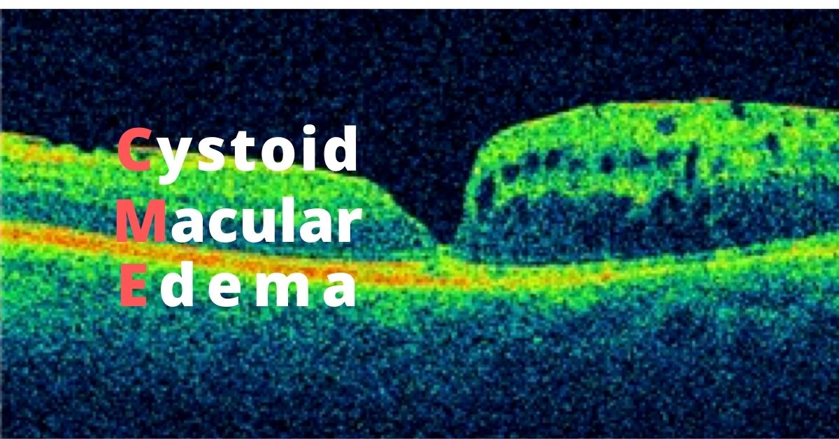
Cystoid Macular Edema

This type of macular swelling is very common and even occurs after cataract surgery.

What is Cystoid Macular Edema?

Cystoid macular edema, aka CME, affects the functional center of the retina, the macula.  It is another term for macular edema where multiple cyst-like areas of swelling develop.

There are multiple causes of CME to include:

  • Retinal Vascular Occlusions (RVO)
  • Intraocular inflammation (uveitis or iritis)
  • Diabetic retinopathy
  • Uncomplicated cataract surgery

Symptoms of Cystoid Macular Edema

The main symptom of CME is blurry vision.  There may also be some symptoms of distortion, poor color perception and minification, but most patients note blurry vision.

Because this is a condition of the macula, only central vision is affected.  The peripheral or side vision is not involved.

Blurry vision can occur from a variety of eye conditions and is not a specific symptom of cystoid macular edema. 

CME after Cataract Surgery

About 1-3% of patients will develop cystoid macular edema following cataract surgery.  This can occur several weeks following uncomplicated cataract surgery.  Before modern phacoemulsification techniques evolved, CME developed in as many as 10% of patients.

For reasons unknown, fluid starts to accumulate in the macula.  Patients are often frustrated because the vision following surgery was excellent, only to get worse. 

Treatment and Prognosis

CME following cataract surgery is usually self limited, that is, there is full restoration of vision and function after several weeks of treatment.  The condition does not usually recur, but there is increased risk of developing CME in the fellow eye.

Treatment for CME following cataract surgery often involves topical anti-inflammatory eye drops.  Steroid drops are often quite effective.  In more complicated cases, injections of steroid around the eye may be helpful

Because CME can develop from other situations other than cataract surgery, treatments and prognosis vary widely.  For instance, cystoid macular edema following a branch retinal vein occlusion may involve anti-VEGF injections or depot steroids.  

Cystoid macular edema can also be caused by certain glaucoma drops or following complicated cataract surgery.  Surgery may be necessary to treat the macular swelling due to complications of cataract surgery. 

Consultation with a retinal specialist may be needed.

Diabetes Awareness: A Look Back And Ahead

November is diabetes awareness month. Read this article to raise your own diabetes aware and share it with others.

November is Diabetes Awareness Month and World Diabetes Day is November 14, the birthday of the man credited with the discovery of insulin—Frederick Banting.

Diabetes is a chronic, metabolic disease which can lead, over time, to serious damage to the heart, blood vessels, eyes, kidneys, and nerves. Diabetes awareness aims to improve understanding of this disease within your family and your community.

An estimated 34.3 million Americans, 10.5%, have diabetes according to the CDC’s National Diabetes Statistics Report, 2020 and 88 million adults, approximately 1 in 3, have prediabetes. New diagnosed cases for both type 1 and type 2 diabetes are on the rise for U.S. youths.  

Diabetic retinopathy, the disease that affects the eyes, is a leading cause of blindness, yet vision loss can be prevented.

A Look Back

Fredrick Banting was a Canadian orthopedic surgeon who was fascinated with the published research into the role of the pancreas in diabetes. In 1901 it was discovered that the pancreas produces insulin from a collection of cells known as the Islets of Langerhans, which are  irregularly shaped patches of endocrine tissue located within the pancreas.  

The problem Banting wanted to solve was extracting the insulin from the pancreas. Many attempts had been made, but all of them had failed.

Banting had an idea but had no research experience and needed help. He convinced the head of the University of Toronto to give him a laboratory and a research assistant.  Banting and his research assistant, Charles Best, started work on May 17, 1921. James Collip, an experienced Canadian biochemist, was asked to join Banting and Best in December 1921 to purify their insulin extract.  

On January 23, 1922, a 14-year-old boy, Lenard Thompson, became the first person to receive an insulin injection as treatment for diabetes. By 1923 insulin was widely available and a diagnosis of diabetes was no longer an immediate death sentence.

Insulin from cattle and pigs was used for many years, but it could cause allergic reactions in some patients. In 1978 recombinant DNA technology made it possible to insert a human gene into a common bacterium so it will produce the protein (insulin) encoded by the human gene. The insulin is extracted from the bacteria and purified.

Today and a Look Ahead

Diabetes is now a manageable disease and the technology for managing it is constantly evolving.  The American Diabetes Association (ADA) has a Device Technology section on its website that describes and explains continuous glucose monitoring devices, glucose meters, and insulin pumps.

On the ADA website you also can find a Diabetes Education Program. These programs are located throughout the U.S. and the educators in the programs work with diabetics to give them the information, skills, and resources they need to successfully manage their diabetes. Medicare and most insurance plans cover these diabetes education programs.

On September 28, 2016 the FDA approved the first artificial pancreas device system. It works by inserting a very tiny sensor under the skin, usually on the stomach or arm, that measures the glucose fluid in the fluid between cells every few minutes and sends this information to an insulin pump attached to the body that, when needed, delivers the right amount of insulin. In this way insulin is released throughout the day as a natural pancreas would.

Islet cell transplants are in the experimental phase and being done in 17 centers around the country that are participating in islet cell research programs. The challenges the researcher face are methods for collecting enough islet cells for a transplant and preventing rejection of the transplant.

What Can You Do?

Improve your own diabetes awareness.

Living with diabetes is now easier than ever before. Share this information with your friends and family to raise their level of knowledge and understanding of this disease.

No matter what, every patient with diabetes needs to have their eyes examined yearly. Severe vision loss and blindness can be prevented, but only with timely and regular examination.

If you would like to schedule an appointment, please call us (877) 245.2020.
Nader Moinfar, M.D., M.P.H.
Retina Specialist
Orlando, FL

Eating Carrots to Improve Your Eye Health

Do carrots really help your vision? Here’s how carrots improve your eye health.

Getting enough vitamin A is essential to maintain eye health and carrots are an excellent natural source of vitamin A, but eating a diet overloaded with carrots or other sources of vitamin A won’t improve your eyesight.

Eye health and good eyesight are different things. Healthy eyes can still need glasses to clearly focus on near or far objects because the shape of the cornea or the eyeball causes nearsightedness, farsightedness, or astigmatism.  No amount of vitamin A will change that.

Vitamin A and Eye Health

Vitamin A is converted into rhodopsin, the photopigment in the rods of your retina that enable vision in low-light. In humans and many animals rhodopsin is required for vision in dim light. 

Rhodopsin converts light into electrical nerve signals within the photoreceptor rods and those electrical signals are sent to the brain via the optic nerve. 

Vitamin A also supports the healthy functioning of the eye’s conjunctival membranes and the corneas.

Vitamin A deficiencies are rare in the U.S. and other developed countries. Most diets in developed countries contain adequate amounts of vitamin A and vitamin A can be stored by the body in the liver. 

Excellent Sources of Vitamin A

Sweet potatoes, spinach, pumpkin, carrots, milk, cheese, cantaloupe, mangos, eggs, and sweet red peppers are all excellent sources of vitamin A.

Vitamins and Oxidative Stress

The human body produces oxygen-derived free radicals as part of the metabolic process, part of breathing and burning energy, and free radicals are essential to life, but their number must be balanced out with antioxidants for optimal health.

A diet rich in antioxidants such as vitamins A, E, and carotenoids such as beta carotene, lycopene, and lutein will help your body scavenge free radicals, terminating their chain reactions before they cause cellular damage.

There is a peptide (glutathione) and some antioxidant enzymes produced by the body specifically to scavenge free radicals, but consuming a diet of antioxidant-rich fruits and vegetables, will also be a diet low in saturated fat and cholesterol, so eating lots of high-antioxidant fruits and vegetables will not only add to your body’s antioxidant levels, but give your overall health a boost.  

If you would like to schedule an appointment, please call us (877) 245.2020.

Nader Moinfar, M.D., M.P.H.
Retina Specialist
Orlando, FL

Drug Delivery for Eye Disease

Eye drops are great for only certain eye diseases. There are other routes used to treat your retina.

There are different routes of drug delivery to treat your eye disease.

Not all parts of the eye can be reached by the same route. Eye drops and ointments applied by patients directly to the eye is the route to get to the cornea, conjunctiva, and sclera, but getting to the retina in the back of the eye requires a different route—an injection.

Intravitreal Injections

Only an intravitreal injection will reach the retina because the retina is part of the brain and is protected by the blood-brain barrier.

Intravitreal means that the injection is into the vitreous of the eye. The vitreous is a clear, colorless, jelly-like substance that fills the space between the lens and the retina. Intravitreal injections of anti-VEGF are placed in the vitreous near the retina to treat wet macular degeneration and diabetic retinopathy.

The injections are done in a doctor’s office and the eye is numbed before the injection. They are a safe and effective way to prevent further vision loss.

Eye Drops

Eye drops are the preferred route of drug delivery to treat eye diseases in the front part of the eye. They are commonly used to treat eye infections, allergies, inflammation, glaucoma, and to provide lubrication for dry eyes.

Eye drops come in solutions and suspensions. In a solution the particle sizes are very small and they completely dissolve in the solvent mixture making a solution clear. Suspensions consist of larger particles that are suspended in a solvent. The larger particles will settle to the bottom so suspensions must be shaken before use to re-suspend the therapeutic particles.

If you are applying more than one drop to an eye, allow the first drop to absorb completely before applying a second drop so the medication doesn’t end up running down your cheek.

Medicated Contact Lenses

For some corneal infections a special type of contact lens or a collagen shield is soaked in antibiotics and placed on the eye.

Ointments

Although ointments are greasy and somewhat difficult to apply they are the preferred eye medication for use on infants and very small children because they stay in the eye longer than eye drops. Ointments are also used for nighttime applications of eye medication so the ointment can cover the eye while you sleep.

Sustained Release Drug Systems

These are biodegradable, implanted devices that supply a slow, steady release of a drug over an extended time to maintain the therapeutic level of the drug to the target area. It is new technology and is available for the treatment of glaucoma, vascular occlusions and diabetic retinopathy.

If you would like to schedule an appointment, please call us (877) 245.2020.

Nader Moinfar, M.D., M.P.H.
Retina Specialist
Orlando, FL

Why Injections Not Drops

Here’s why eye drops don’t work for treating diseases of the retina.

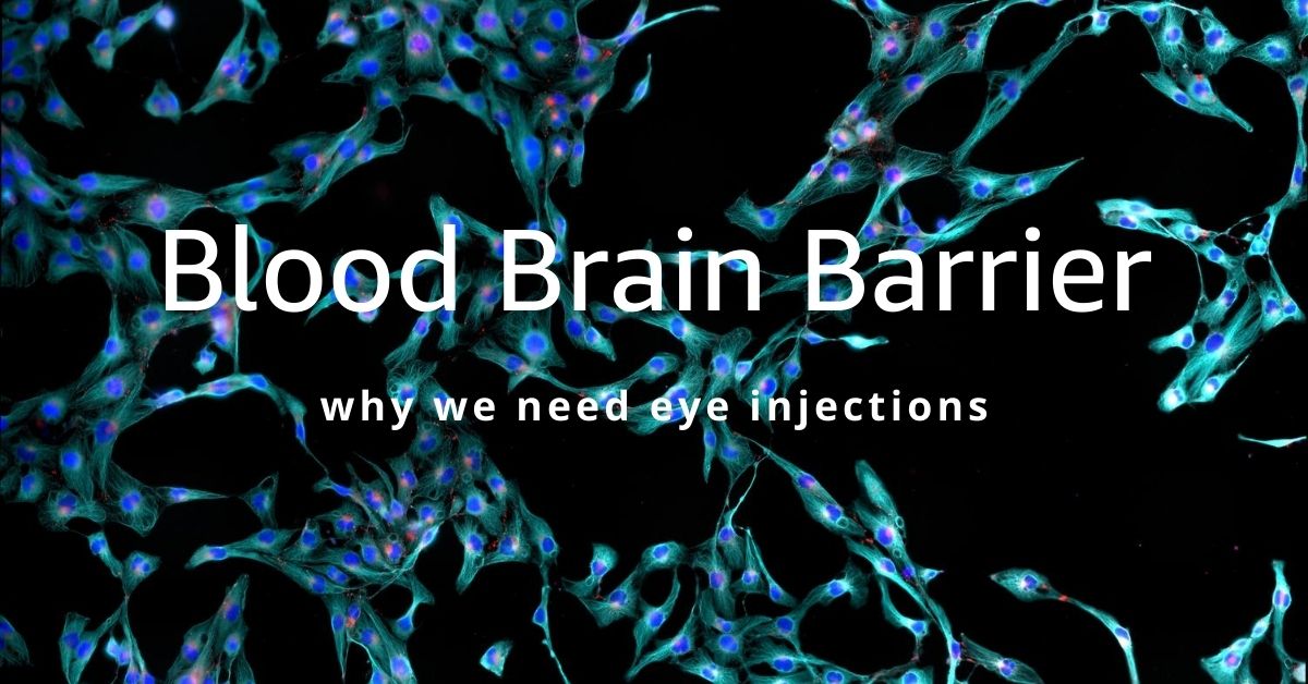
The blood brain barrier blocks most medications from reaching the retina. You may wonder why there isn’t a pill, or an eye drop that treats these retinal diseases and why the only effective treatment is a shot in the eye, an intraocular injection.

Intraocular injections are common and effective ways to treat wet macular degeneration, diabetic retinopathy and retinal vascular occlusions.

Pills, eye drops, and even IV solutions cannot penetrate to the retina because it is protected by the blood-brain barrier. Direct injection circumvents this barrier.

The eye is the only part of the brain that can be viewed directly. And that is done when an eye doctor uses an ophthalmoscope and shines a bright light into your eye and can view the innermost layers of the eye—the retina, and the optic nerve.

Blood Brain Barrier

The blood-brain barrier (BBB) is a protective layer of endothelial cells that protects the brain from any pathogens or toxins that may be circulating in the blood supply, but it also restricts entry of large-molecule medications and 98% of all small-molecule drugs. So, the only way to get therapeutics to the brain or to the deep structures of the eye is to go through or behind the BBB.

The BBB allows water, some gasses, and fat-soluble molecules to dissolve in the cell membranes and cross its boundary. Alcohol, caffeine, and nicotine easily cross the BBB because they are lipophilic (attracted to fat) and mix easily with the fat in the BBB to gain entry. Oxygen and anesthesia are gasses that can pass through the BBB.

As part of the brain, the BBB prevents therapeutics that have been ingested and are circulating in the blood to gain entry to the eyes. The medication will travel through the body, but almost none of it will reach the eyes.

Leaky Blood Vessels

Wet macular degeneration is caused when blood vessels behind the retina grow abnormally and leak blood and fluid that distorts vision. If left untreated the leaking blood vessels will cause irreversible damage to the photoreceptors that create central vision.

A Shot in the Eye

The only way to get enough medication directly to the retina to stop the growth of the leaky blood vessels is to use an intravitreal injection. An intravitreal injection means that the medication is injected into the vitreous (jelly-like fluid) of the eye where it will diffuse to the retina.

In addition, injecting anti-vascular endothelial growth factor (anti-VEGF) directly into the eye where the leaky vessels are growing, prevents the anti-VEGF from circulating in your blood supply and possibly adversely affecting systemic blood vessels as it moves along to the eye.

The intravitreal injections are painless because the eye is numbed, and the injections can be done in your doctor’s office. They are a safe, and effective way to prevent leaky blood vessel damage to your retina.

Coming Advances in Treatment

An implanted reservoir that delivers sustained-release anti-VEGF directly to the retina is in the experimental phase. And the field of nanotechnology is in the preliminary research stages of developing nanoparticles as a potential way to transfer medication in eye drops across the BBB.

If you would like to schedule an appointment, please call us (877) 245.2020.

Nader Moinfar, M.D., M.P.H.
Retina Specialist
Orlando, FL

Symptoms of a PVD

These symptoms may prevent a retinal detachment and here’s why.

PVD stands for “posterior vitreous detachment.” It’s a normal event that increases your risk for developing a retinal tear which can lead to a retinal detachment. Everyone will eventually get a PVD.

New flashes and new floaters should be reported to your eye doctor or retina specialist. Not all floaters are benign. Only a full dilated exam will determine if your new symptoms are related to a posterior vitreous detachment, retinal tear or something else.

Symptoms of Posterior Vitreous Detachment

The most common symptoms of a PVD:

  • Flashes
  • Floaters
  • Nothing (Asymptomatic, no flashes or floaters)

There are two ways to stimulate the retina: normal light and physical manipulation. Usually, light enters the eye and is captured by the rods and cones of the retina which eventually produce vision. The other mechanism occurs when you rub your eyes so hard you actually see patterns in your vision. This rubbing, or physical manipulation, also produces “light.”

After a PVD has occurred, only the back, or posterior portion, of the vitreous separates from the retina. The anterior portion remains fixed to the surface of the retina. As the newly separated vitreous cleaves from the retinal surface in the back, it can move to and fro in the eye. This transmits energy to the portion of the vitreous still attached to the retina in the anterior part of the eye…causing flashes.

What are Floaters?

Floaters can be a result of cellular debris from underneath the retina in cases of a retinal tear, blood or just opacities which have formed in the otherwise clear vitreous gel.

Not all posterior vitreous detachments cause flashes or floaters. Thus, follow up is always recommended so your retina specialist can look for asymptomatic retinal tears.

What if I have a Retinal Tear?

First of all new flashes and floaters should be reported to your eye doctor. In the case of a PVD, it’s important to look for a retinal tear. A retinal tear is most likely to occur during the first 6 weeks after a PVD has occurred.

The goal of examination is to find a retinal tear before it turns into a retinal detachment. Retinal tears can usually be treated with laser whereas a retinal detachment requires surgery and there is a chance of lost vision.

If you would like to schedule an appointment, please call us (877) 245.2020.
Nader Moinfar, M.D., M.P.H.
Retina Specialist
Orlando, FL

What is Legal Blindness?

Legal blindness can be caused by a variety of diseases. Here’s how it is defined and links to resources.

Legal blindness is a legal definition that is used to refer to patients with vision loss who qualify for government assistance. This is not total blindness.

Legal blindness is defined as a patient’s best visual acuity (aka best corrected) in either eye is 20/200 or less or the size of the visual field (peripheral vision) is less than 20 degrees. In other words, if both eyes do not see well, the vision is no better than 20/200 in better seeing eye.

“Best corrected” refers to the use of the best corrective lens/contact lens for the patient.

A central visual acuity of 20/200 or worse means that a legally blind person must be 20 feet away from an object to see what a “normal” person can see from 200 feet away.

Loss of peripheral vision can also qualify as legal blindness. Most patients normal peripheral vision can see at least 140 degrees without turning the head. Legally blind peripheral vision is less than 20 degrees.

Causes of Legal Blindness

  • Age-related macular degeneration (ARMD) is one of the leading causes of legal blindness in Americans aged 60 and older. ARMD affects the macula (functional center of the retina) and therefore decreases central vision.
  • Cataracts will affect almost everyone by the age of 80. Cataracts blur both central and peripheral vision (though reversible with cataract surgery).
  • Diabetic retinopathy is another leading cause of blindness and affects the blood vessels in the back of the retina. Diabetic retinopathy causes either blurring of the central vision (most common), but can affect peripheral vision, too.
  • Glaucoma is a progressive disease that damages the optic nerve. Glaucoma primarily causes visual field loss and, only in the late stages, affects central vision.

NOTE: All of the disease described above have available treatments which, in most cases, can preserve vision or slow down progression if diagnosed timely.

Low Vision Aids

May low-vision aids and devices are available to assist individuals who are legally blind. Both near and distance vision can be improved with low-vision aids.

For example, desktop, stand-alone and hand-held magnifiers are available. These may be helpful for close range work such as reading or computer use. Prices vary up to several hundred dollars.

Wearable devices that magnify are also available. Mounted binoculars, wearable HD autofocus cameras with TV viewing and electronic headsets with built-in cameras can help patients with central and peripheral vision loss. Prices vary up to several thousand dollars.

Resources

There are many governmental and non-governmental resources for those who are legally blind.

For example, the American Foundation for the Blind (AFB) can assist those with low-vision. Founded in 1921, the AFB ensures that patients who are blind, legally blind or otherwise visually impaired have access to educational materails, technology and legal information.

Here are additional resources that help blind, legal blindness and visually impaired.

Low Vision Evaluation

If you feel you suffer from legal blindness or need more information, a low-vision examination is the first place to start. Eye doctors specializing in low vision can advise and educate you about the best low-vision aids for your specific visual needs.

If you would like to schedule an appointment, please call us (877) 245.2020.

Nader Moinfar, M.D., M.P.H.
Retina Specialist
Orlando, FL

Possible COVID-19 Treatment can Cause Eye Damage

Hydroxychloroquine is FDA approved for certain autoimmune diseases and malaria, but has the potential for toxicity…

Hydroxychloroquine has long been used to safely treat patients with malaria and certain autoimmune diseases such as lupus erythematosus and rheumatoid arthritis, but NOT COVID019.

In the news, there have been small, inconclusive studies using two malaria and autoimmune medications to treat COVID-19 (coronavirus 2019). Some studies suggest the medications might be helpful, while other studies saw no difference.

Currently, there is not enough medical data to prove that hydroxychloroquine and chloroquine work for COVID-19.

Hydroxychloroquine Approved for Malaria

Chloroquine was approved by the FDA in 1949 as a treatment for malaria, an infection caused by a parasite. Hydroxychloroquine was approved by the FDA in 1955 for the treatment of malaria. Hydroxychloroquine is preferred over chloroquine because it has fewer side effects.

Both drugs are available in pill form. Plaquenil is also approved for long-term use in rheumatoid arthritis and lupus.

The side effects of both medications include:

  • Retinal damage and irreversible vision loss (Plaquenil Toxicity)
  • Headache
  • Abnormal heart rhythm
  • Muscle weakness or nerve pain
  • Gastrointestinal issues (nausea, cramping, vomiting, and diarrhea)
  • Hypoglycemia (low blood glucose)
  • Worsening of psoriasis

Retinal Damage | Plaquenil Toxicity

The overall prevalence of chloroquine/hydroxychloroquine retinopathy is 7.5%. Retinal damage is one of the most serious adverse effects of hydroxychloroquine (HCQ) since the retinal damage caused by the drug is irreversible. Chloroquine/hydroxychloroquine retinopathy is more common with higher daily doses and long-term use.

A baseline retinal examination is recommended for all patients taking HCQ, along with regular monitoring examinations. The baseline and monitoring examinations should include the following:

  • Uncorrected visual acuity test
  • Best-corrected visual acuity test
  • Examination with ophthalmoscope
  • Examination using slit-lamp
  • Optical Coherence Tomography

The FDA previously issued an Emergency Use Authorization (EUA) that allows physicians to request a supply of hydroxychloroquine or chloroquine for hospitalized patients with COVID-19 who are unable to join a clinical trial. 

As of this writing 4/24/20, the FDA now warns against the use of hydroxychloroquine for the treatment of COVID-19 after reports of irregular heart rhythms and deaths.

If you are presently taking Plaquenil, talk to your prescribing doctor to insure that you are getting regular eye exams.

If you would like to schedule an appointment, please call us (877) 245.2020.

Nader Moinfar, M.D., M.P.H.
Retina Specialist
Orlando, FL

Symptoms of Eye Emergencies

During the pandemic, the American Academy of Ophthalmology and the Centers for Disease Control and Prevention, have mandated that your eye doctor limit practice to those patients with eye emergencies or urgencies. Elective surgery has been postponed.

There are several warning signs or symptoms which should alert you to call your eye doctor.

Pain

Eye pain can be caused by a corneal abrasion, inflammation or advanced glaucoma.

Corneal abrasions may also be associated with tearing, redness and blurry vision. The corneal is ultra sensitive and even a slight abrasion can be painful. The corneal surface allows for about 2/3 of the total focusing power of the eye. If the surface becomes abraded, blurred vision is common.

Inflammation, such as iritis, causes pain, redness, sensitivity to light and blurry vision. Advanced glaucoma can lead to uncontrolled eye pressure causing pain, redness, tearing, nausea and decreased vision.

Eye pain associated with redness should prompt a call to your doctor. Almost any and every “eye related” cause of eye pain involves redness.

Sudden Decreased Vision

Sudden decreased or vision loss can be an emergency.

Most retinal diseases can cause sudden and painless vision loss, such as:

While there are other causes, I’m listing the common causes of acute vision loss from retinal diseases.

Sudden Distortion

Distortion is usually caused by a macular problem.

While distortion is a common complaint in patients with macular holes or epiretinal membranes, the onset is usually gradual and not acute.

Acute distortion can be from macular edema (swelling) from an retinal vascular occlusion or leakage/bleeding from wet macular degeneration. Sometimes a retinal detachment can cause distortion, but this is usually associated with complaints of loss of peripheral vision.

Exacerbation of wet macular degeneration is certainly an emergency and may require prompt treatment with anti-VEGF medications.

If you are experiencing pain, loss of vision or new distortion, make sure to call your eye doctor or retina specialist.

If you would like to schedule an appointment, please call us (877) 245.2020.

Nader Moinfar, M.D., M.P.H.
Retina Specialist
Orlando, FL

Jon Doe