Dislocated Intraocular Lens

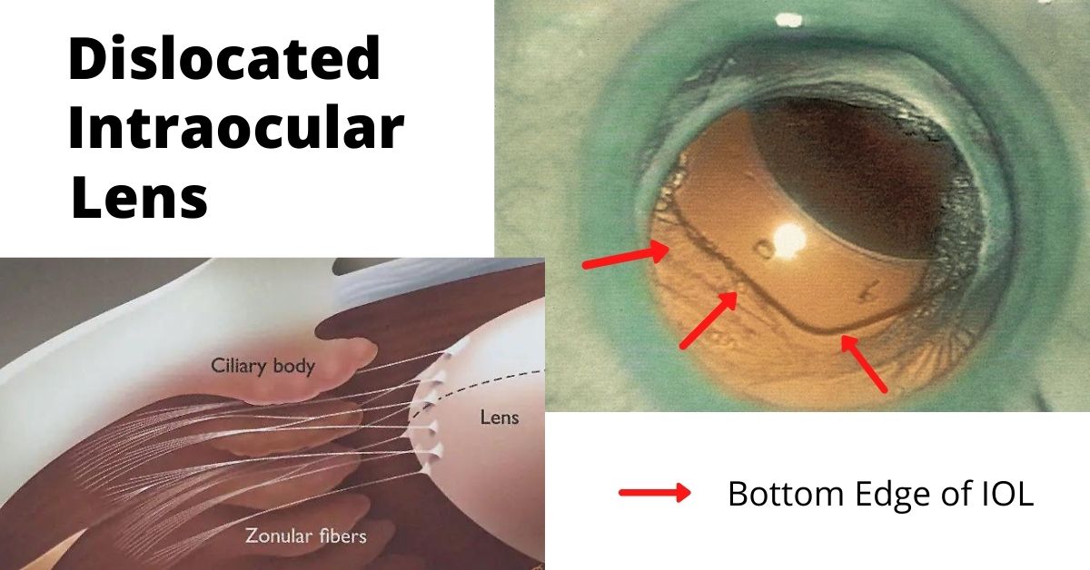
A dislocated intraocular lens is very similar to a dislocated crystalline (natural) lens. The only difference is that the intraocular lens has been inserted during previous cataract surgery.

Cataract surgery is a low-risk surgery and has a success rate of about 95%. But like all surgeries there can be complications, especially if you have other eye or health issues.  

One of those complications is a dislocated intraocular lens (IOL). This is a rare complication and occurs in less than 3% of cases, but it may require additional surgery to repair.

What causes a Dislocated?

Sometimes the dislocation happens shortly after cataract surgery because the zonules, a ring of tiny thread-like fibers that support and hold the lens capsule in place, fail or break. The zonules are made of a connective tissue protein. People who have a connective tissue disorder are more likely to have this complication, and it can also occur in patients who have a history of previous eye surgeries, trauma to the eye, or who take prostate medication.

Other times the IOL dislocation happens gradually over time when the zonules slowly weaken and don’t adequately support the lens capsule and this causes the lens to shift away from the center of the pupil. This happens more often in patients with a history of eye trauma, multiple eye surgeries, especially retinal detachment repair, and complicated original cataract surgery.

Symptoms

The most common symptom of a dislocated IOL is blurry vision. The degree of blurriness depends on how dislocated the IOL is. If the IOL is only slightly dislocated then vision may still be clear, but ghost images or a double image at night may occur when light passes through the edge of the dislocated lens.

Treatments

Treatment depends on the severity of the dislocation. Some dislocations very minor and vision is still good. In these cases, no treatment is necessary.  

If the lens is completely off center and vision is very blurry, then only surgery will correct it. In some cases, the original implanted IOL can be repositioned and secured in place. In other cases when the zonules are extremely weak or damaged, a new IOL designed to be sutured to the wall of the eye (sclera) or to the iris is used.

If the lens has fallen into the vitreous cavity, then a retinal surgeon will need to perform a vitrectomy to remove the vitreous jelly inside the eye and retrieve the dislocated intraocular lens.  

Prognosis

Most patients who have IOL repositioning or IOL replacement have vision of 20/40 or better after surgery. The outcome depends on your health and if you have other eye issues, such as macular degeneration or diabetic retinopathy.  

Recovery for IOL replacement is similar to the recovery time for cataract surgery. The medications are the same and most patients can resume normal activities the day after surgery.

If you would like to schedule an appointment, please call us (877) 245.2020.
Nader Moinfar, M.D., M.P.H.
Retina Specialist
Orlando, FL

Dislocation of the Natural Lens

The fancy term for diplacement of your natural lens in the eye is called ectopia lentis…here’s when it happens.

Your natural lens can become displaced from it’s natural anatomic position within the eye. Intraocular lenses can also become displaced, but this article is limited to dislocation of the natural crystalline lens.

Ectopia lentis is the medical term for the dislocation or displacement of the eye’s natural crystalline lens. The lens may be free-floating in the vitreous or it may be in the anterior chamber or directly on the retina.

This dislocation most commonly occurs after trauma to the eye. It can also be caused by a systemic disease, such as Marfan syndrome. Trauma is the most common cause and most often the result of a direct blow to the eye such as from a baseball or golf ball.

Some rare and inherited genetic diseases can cause ectopia lentis:

  • Weill-Marchesani syndrome
  • Sulfite oxidase deficiency
  • Hyperlysinemia
  • Ehlers-Danlos syndrome
  • Sturge-Weber syndrome
  • Mandibulofacial dysostosis
  • Wildervanck syndrome
  • Conradi syndrome
  • Pfaundler syndrome
  • Crouzon syndrome
  • Pierre Robin syndrome
  • Sprengel deformity

Symptoms of Lens Dislocation

The most significant symptom of ectopia lentis is reduced visual acuity such as poor near or distant vision. The degree of reduction in visual acuity varies with the degree of lens dislocation and the type of dislocation.

If the zonules in the eye are disrupted it can lead to increased curvature of the lens and may result in lenticular myopia or astigmatism. The zonules are tiny thread-like fibers that hold the eye’s lens firmly in place and also tighten the pull the lens to accommodate near vision.

Treatment

Treatment of ectopia lentis depends on where and how far the lens has moved and any resulting complications. In some cases, in which the dislocation is minimal and there is no significant impact on vision observation and close follow-up are the only treatments necessary.

In cases in which vision is affected or there is damage to surrounding structures, surgery may be necessary. The dislocated lens is removed, and an artificial lens is put into place. If there is not enough structural support for the artificial lens then it may need to be sutured to the iris or sclera.

Prognosis

Multiple surgical techniques are available for correction of ectopia lentis and each has its own limitations and associated complications. In some cases, an implanted artificial lens is used, and other patients are treated by removal of the lens and a vitrectomy to remove the vitreous humor and then they use special contact lenses.  

If the lens dislocated due to a genetic disease then the underlying disease must also be treated.

Most patients with ectopia lentis do well and at least 85% achieve a 20/40 or better visual acuity. However, if there is a pre-existing condition such as a corneal disease, glaucoma, diabetic retinopathy, macular degeneration, or a history of retinal detachment, the outcome will not be as favorable.

Close follow-up is essential as is medical management of any complications that may arise. 

If you would like to schedule an appointment, please call us (877) 245.2020.

Nader Moinfar, M.D., M.P.H.
Retina Specialist
Orlando, FL

Jon Doe