What is Macular Pucker?

Macular pucker, also known as an epiretinal membrane or cellophane maculopathy, is a condition that affects the macula, a small area located in the center of the retina at the back of the eye. The macula is responsible for central vision, which allows us to see fine details and perform tasks such as reading, driving, and recognizing faces.

Causes of Macular Pucker


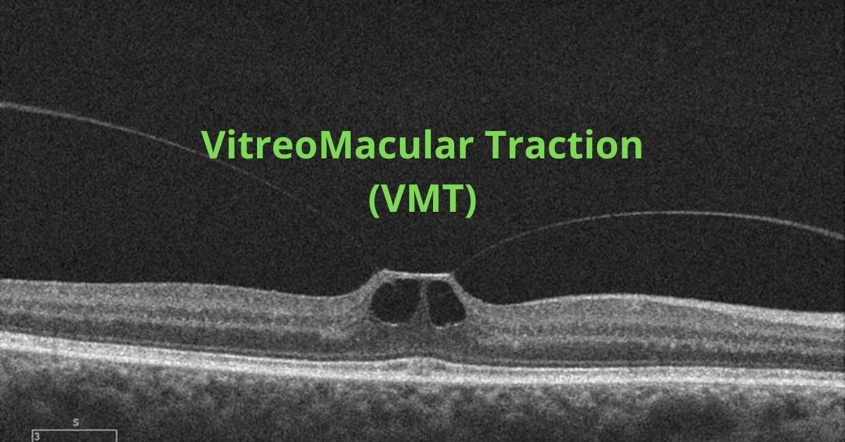
Macular pucker occurs when a thin, transparent layer of scar tissue forms on the surface of the macula. This scar tissue can cause the macula to wrinkle or pucker, leading to distorted and blurred vision. The most common cause of epiretinal membrane development is aging, as the vitreous gel inside the eye shrinks and pulls on the macula, causing the formation of scar tissue. However, other factors such as eye trauma, inflammation, and certain eye conditions can also contribute to its development.

Symptoms of Macular Pucker


Symptoms of macular pucker can vary from mild to severe and typically affect only one eye. Common symptoms include blurred or distorted central vision, difficulty reading or performing tasks that require good vision, and the appearance of straight lines as wavy or bent. In some cases, individuals may also experience a gray or cloudy area in their central vision.


While macular pucker can cause significant visual impairment, it can vary in severity. In mild cases, no treatment may be necessary, and individuals can adapt to their vision changes. However, in more severe cases where vision loss significantly affects daily activities, surgical intervention may be recommended.

Treatment Options


The most common surgical procedure for macular pucker is called vitrectomy with membrane peeling. During this procedure, the vitreous is removed from the eye and the scar tissue on the macula is carefully peeled away. Following surgery, visual recovery may take several weeks to months, but not all individuals will regain normal vision.


Regular eye examinations are important for the early detection of macular pucker or other eye conditions. If you experience any changes in your vision, I recommend you inform your own doctor to provide a proper diagnosis and recommend appropriate treatment options after examining you.

If you would like to schedule an appointment, please call us (877) 245.2020.

Nader Moinfar, M.D., M.P.H.
Retina Specialist
Orlando, FL

Syfovre™ for Geographic Atrophy

Syfovre™ is the first available FDA approved treatment for geographic atrophy, an advanced form of macular degeneration.

Syfovre™ is a novel treatment for an advanced stage macular degeneration called geographic atrophy.

The FDA has approved Syfovre™ (pegcetocplan) for the treatment of GA, a leading cause of in developed countries. Syfovre™ is the first treatment for this advanced stage of macular degeneration. Treatment for wet macular degeneration has been available for over a decade.

Administration of Syfovre™

As with many retinal therpies, Syfovre™ is administered as an intravitreal injection into the eye. Because eye drops, ointments, oral meds and intravenous medications have difficulty penetrating the eye, intravitreal injections are necessary to deliver the right amount of drug to the retina.

Injections may be given monthly or every other month and will need to be repeated. The exact frequency of dosing will be determined by your doctor.

How Does Syfovre™ Work?

Pegcetocoplan is a unique inhibitor of C3, a protein involved in the complement system. The complement system is a major factor in our immune system and helps prevent infection from bacteria, viruses, regulates inflammation and aids the body get rid other pathogens.

Left unchecked, C3 may be involved in the death of healthy cells of the retina called retinal pigment epithelial (RPE) cells. RPE cells are responsible for the health and maintenance of photoreceptors, such as rods and cones. Without healthy photoreceptors, we can not see.

Syfovre™ binds C3 much like an antibody and prevents C3 from causing cell death to the retinal RPE cells.

Geographic Atrophy

Areas of RPE cell loss can be detected during a retinal examination. Usually these so-called “atrophic lesions” arise near the macula. With time the lesions enlarge to involve the central macula.

Visual acuity, your central vision, is usually spared early in the disease, but visual function may be compromised depending upon the size, number and location with respect to the central macula.

Unlike wet macular degeneration where abnormal blood vessels develop within the layers of the retina, geographic atrophy is causes by death of the RPE cells.

Symptoms of Geographic Atrophy

Symptoms of macular degeneration include: blurry vision, distortion and loss of central vision. Geographic atrophy can also cause decline in visual function such as poor contrast, decreased reading speed, poor color perception etc.

If you or your family member is concerned, please call your eye care provider. The earlier this is detected and possibly treated, the better.

If you would like to schedule an appointment, please call us (877) 245.2020.

Nader Moinfar, M.D., M.P.H.
Retina Specialist
Orlando, FL

Treatment of Diabetic Macular Edema (DME)

Diabetic macular edema is a common complication of diabetic retinopathy where fluid accumulates in the macula and blurs the vision.

Diabetic macular edema (DME) is a condition characterized by the accumulation of fluid in the macula, the central part of the retina responsible for sharp, detailed vision. It is a common complication of diabetic retinopathy, a disease affecting the blood vessels of the retina. The management of diabetic macular edema (DME) involves various treatment approaches aimed at reducing macular edema and preserving vision.

Anti-VEGF Injections – Treatment of Diabetic Macular Edema

One of the primary treatment options for DME is intravitreal injections of anti-vascular endothelial growth factor (anti-VEGF) medications. Drugs like ranibizumab, bevacizumab, and aflibercept are commonly used to inhibit the abnormal growth of blood vessels and reduce leakage, thus decreasing macular edema. These injections are typically administered on a monthly or as-needed basis, with the aim of improving visual acuity and reducing central retinal thickness.

Steroids for Diabetic Macular Edema

Another treatment modality for DME is the use of corticosteroids. Intravitreal injections of triamcinolone acetonide or dexamethasone implants can help suppress inflammation and reduce fluid accumulation in the macula. These steroid injections are usually performed at longer intervals compared to anti-VEGF agents due to their sustained-release properties.

In some cases, laser photocoagulation may be employed to treat DME. Focal laser treatment can be used to seal leaking blood vessels and reduce fluid accumulation. However, this approach is typically reserved for cases where the edema is away from the central macula to avoid damaging central vision.

New Treatments

Recently, a new class of medications called integrin receptor antagonists has shown promise in the treatment of DME. These drugs, such as pegpleranib and abicipar, target specific proteins involved in the pathogenesis of macular edema and are administered via intravitreal injections.

In addition to these specific treatments, managing the underlying diabetes is crucial in preventing and managing DME. Maintaining optimal blood sugar control, blood pressure, and cholesterol levels can help reduce the risk and progression of DME.

Regular eye examinations and early detection of DME are essential for timely intervention and optimal outcomes. Treatment options should be individualized based on the severity of the edema, visual impairment, and patient characteristics. By employing a combination of these treatment modalities and a comprehensive approach to diabetes management, healthcare professionals can improve visual outcomes and enhance the quality of life for individuals living with DME.

If you would like to schedule an appointment, please call us (877) 245.2020.

Nader Moinfar, M.D., M.P.H.
Retina Specialist
Orlando, FL

7 Reasons You Need a Retina Specialist

A retina specialist has undergone expert training to deal with diseases and surgery of the retina and vitreous such as these disorders or situations.

A retina specialist is an ophthalmologist who has extra-training in diseases and surgery of the retina. Additional training years are required to focus attention in this special area.

Here are 7 more common reasons why you may be referred to a retina specialist.

Retinal Tear, Retinal Detachment, Retina Specialist

Retinal tears can cause a retinal detachment. Retinal tears may be asymptomatic, but often are associated with the sudden onset of new floaters or flashes usually due to a posterior vitreous detachment.

While most new floaters and flashes are NOT associated with a retinal tear, the only way to make sure you are not at risk for developing a retinal detachment is have a complete dilated eye exam.

Retinal tears can be treated with either cryotherapy or laser. Retinal detachments will often require surgery.

Foreign Body Inside the Eye

While not commonplace, intraocular foreign bodies may occur with any penetrating eye injury. Glass, metals and organic material need to be removed by performing a vitrectomy, the principal operation performed by a retina specialist.

Retina Specialist and Endophthalmitis

Endophthalmitis is infection inside the eye. This usually occurs following recent eye surgery, but can also follow penetrating eye injury.

Prompt referral to a retina specialist is paramount in preventing loss of vision from infection. Intraocular antibiotics, anti-fungals and/or vitrectomy surgery may be necessary to prevent catastrophic vision loss.

Advanced Diabetic Retinopathy

Diabetic retinopathy can cause blurry vision and blindness. A retina specialist is adept at treating the two most common causes of vision loss from diabetes: diabetic macular edema and proliferative diabetic retinopathy.

Common treatments used by retina specialists include: anti-VEFG injections, steroids, laser and surgery. In most scenarios, these are procedures only offered by a retina specialist.

Macular Degeneration

Macular degeneration (AMD) comes in two forms: dry AMD and wet AMD. The “wet” form is also called neovascular AMD (nAMD).

Both types of macular degeneration affect both eyes and are progressive, but the wet form can steal vision quickly, is associated with leaky abnormal blood vessels and bleeding. Treatment usually involves intravitreal injections of anit-VEGF.

There is presently no treatment for dry macular degeneration approved by the FDA.

Retinal Vascular Occlusions

Periodically the retinal vessels can become occluded or blocked. Both arteries and veins can be blocked retinal vascular occlusions. Vision loss is usually sudden and treatments may be required in an attempt to improve vision and/or to prevent total blindness from complications of the vascular occlusions.

If you would like to schedule an appointment, please call us (877) 245.2020.

Nader Moinfar, M.D., M.P.H.
Retina Specialist
Orlando, FL

Dislocation of the Natural Lens

The fancy term for diplacement of your natural lens in the eye is called ectopia lentis…here’s when it happens.

Your natural lens can become displaced from it’s natural anatomic position within the eye. Intraocular lenses can also become displaced, but this article is limited to dislocation of the natural crystalline lens.

Ectopia lentis is the medical term for the dislocation or displacement of the eye’s natural crystalline lens. The lens may be free-floating in the vitreous or it may be in the anterior chamber or directly on the retina.

This dislocation most commonly occurs after trauma to the eye. It can also be caused by a systemic disease, such as Marfan syndrome. Trauma is the most common cause and most often the result of a direct blow to the eye such as from a baseball or golf ball.

Some rare and inherited genetic diseases can cause ectopia lentis:

  • Weill-Marchesani syndrome
  • Sulfite oxidase deficiency
  • Hyperlysinemia
  • Ehlers-Danlos syndrome
  • Sturge-Weber syndrome
  • Mandibulofacial dysostosis
  • Wildervanck syndrome
  • Conradi syndrome
  • Pfaundler syndrome
  • Crouzon syndrome
  • Pierre Robin syndrome
  • Sprengel deformity

Symptoms of Lens Dislocation

The most significant symptom of ectopia lentis is reduced visual acuity such as poor near or distant vision. The degree of reduction in visual acuity varies with the degree of lens dislocation and the type of dislocation.

If the zonules in the eye are disrupted it can lead to increased curvature of the lens and may result in lenticular myopia or astigmatism. The zonules are tiny thread-like fibers that hold the eye’s lens firmly in place and also tighten the pull the lens to accommodate near vision.

Treatment

Treatment of ectopia lentis depends on where and how far the lens has moved and any resulting complications. In some cases, in which the dislocation is minimal and there is no significant impact on vision observation and close follow-up are the only treatments necessary.

In cases in which vision is affected or there is damage to surrounding structures, surgery may be necessary. The dislocated lens is removed, and an artificial lens is put into place. If there is not enough structural support for the artificial lens then it may need to be sutured to the iris or sclera.

Prognosis

Multiple surgical techniques are available for correction of ectopia lentis and each has its own limitations and associated complications. In some cases, an implanted artificial lens is used, and other patients are treated by removal of the lens and a vitrectomy to remove the vitreous humor and then they use special contact lenses.  

If the lens dislocated due to a genetic disease then the underlying disease must also be treated.

Most patients with ectopia lentis do well and at least 85% achieve a 20/40 or better visual acuity. However, if there is a pre-existing condition such as a corneal disease, glaucoma, diabetic retinopathy, macular degeneration, or a history of retinal detachment, the outcome will not be as favorable.

Close follow-up is essential as is medical management of any complications that may arise. 

If you would like to schedule an appointment, please call us (877) 245.2020.

Nader Moinfar, M.D., M.P.H.
Retina Specialist
Orlando, FL

Vitreomacular Traction Syndrome

VMT is very similar to macular pucker and causes the same changes to your vision.

Vitreomacular traction (VMT) is a complication that arises from a normal process. That normal process is posterior vitreous detachment (PVD) and it happens to everyone as they age. By about age 40 or 50 the vitreous gel that fills the eye begins to change. It begins to shrink and lose fluid and strands of the gel can drift through the eye. These strands can be seen as dark strings or spots that float around in your field of view and are called floaters.

The vitreous gel eventually completely separates from the retina. This is perfectly normal and happens to most people by the age of 70, that is, a PVD is a completely normal event.

Problems arise only if the vitreous gel is strongly attached to the retina, specifically at the macula. If that is the case then when the vitreous gel shrinks it can pull on the retina.

That sticking process is called vitreomacular traction and is very similar to an epiretinal membrane.

The pulling and tugging on the center of the retina where the macula is located can damage the macula and cause vision loss if left untreated.

In healthy eyes, VMT is not common. Certain eye conditions or diseases put people at a higher risk of developing VMT. Those conditions and diseases include:

  • High myopia which is extreme nearsightedness
  • Age-related macular degeneration which is a breakdown of the tissues in the back of the eye
  • Diabetic eye disease which affects the blood vessels in the back of the eye
  • Retinal vein occlusion which is a blockage of veins in the retina

Symptoms

The most common symptoms of VMT include:

  • Distorted vision that makes straight lines appear wavy, blurry or have blank spots
  • Seeing lots of flashes of light in your vision
  • Seeing objects as smaller than their actual size

It is important to see an ophthalmologist for an evaluation when you first notice any of these symptoms.

Diagnosis

To diagnose vitreomacular traction (VMT) your ophthalmologist will use tests such as optical coherence tomography (OCT) which uses light waves to take pictures of the various layers of the retina and will show any damage to the macula.

Fluorescein angiography may also be used. This is an imaging test to view how well the blood is circulating inside the retina and to find any macula swelling. It uses a medical imaging dye injected into the arm that circulates inside the retina while a special camera photographs the progress of the dye as it moves through the blood vessels in the eye.

An ultrasound scan may also be used so your ophthalmologist can get a better view of the location of the sticking point between the vitreous and the macula.

Treatment

Some cases do not require treatment and will resolve on their own, but you will be asked to monitor your vision at home with a grid of lines to make sure the VMT does not progress. If the lines on the grid begin to appear wavy or have missing areas then you will most likely require treatment.

Surgery to remove the vitreous and replace it with a saline solution may be needed to prevent macular holes, puckers or macular swelling from developing or worsening.

Some people are candidates for medication treatments. A medication that dissolves the proteins that link the vitreous to the macula can be injected into the eye. Usually only one injection is needed.

Prognosis

Most patients with VMT maintain good visual acuity in the affected eye even if treatment is required.

If you would like to schedule an appointment, please call us (877) 245.2020.

Nader Moinfar, M.D., M.P.H.
Retina Specialist
Orlando, FL

Eye Scan Detects Alzheimer’s Disease

A simple retina scan may soon become a non-invasive method for diagnosing Alzheimer’s disease, and those at risk.

The abnormal proteins that cause Alzheimer’s Disease can build up in the brain two decades before the onset of symptoms and researchers have been searching for ways to detect the disease sooner. Early detection could mean that medications that slow Alzheimer’s progression could be started sooner. The effectiveness of any early treatments could be monitored with eye scans.

Non-Invasive Hi-Res Imaging

Researchers at Duke University and at Washington University School of Medicine in St. Louis have found evidence that Optical Coherence Tomography (OCT) scans can detect the changes in the tiny capillaries and the thinning of the retinal layers in the eye that signal the beginnings of Alzheimer’s disease.

Dr Sharon Fekrat, from Duke University Medical Centre in the US, said: “We’re measuring blood vessels that can’t be seen during a regular eye exam and we’re doing that with relatively new noninvasive technology that takes high-resolution images of very small blood vessels within the retina in just a few minutes.”

Alzheimer’s Causes Retinal Thinning

Microscopic blood vessels form a dense web at the back of the eye inside the retina. In Alzheimer’s patients, researchers saw that that web was less dense, and there was a loss of small retinal blood vessels. The retinal nerve fiber layer was also thinner in patients without Alzheimer’s disease.

Researchers think that reduced levels of acetylcholine in the brain causes some of the symptoms of Alzheimer’s disease. Acetylcholine is one of the neurotransmitter chemicals that nerve cells use to communicate with one another in the brain. Acetylcholinesterase inhibitors prevent the breakdown of acetylcholine and are used to boost the cell-to-cell communication that gets depleted in Alzheimer’s disease.

What is OCT?

Optical coherence tomography is a non-invasive imaging test that uses light waves to take cross-section images of the retina. An OCT shows each of the distinctive layers of the retina. The OCT produces a three-dimensional map of the eye and can show areas of the eye that are abnormal.

The images from an OCT scan are high resolution because they are based on light, rather than sound, as in an ultrasound, or radio frequency, as in an MRI. Because the OCT shows cross-sections of tissues layers, nerve fiber thickness can be measured. 

If you would like to schedule an appointment, please call us (877) 245.2020.

Nader Moinfar, M.D., M.P.H.
Retina Specialist
Orlando, FL

What Causes a Macular Hole?

Retinal surgery sometimes uses injection of gas into the eye, especially when you have a macular hole.

Causes of a macular hole are discussed in this article and explained in the embedded video.

A macular hole is a hole at the very center of the retina. The retina is the layer of tissue that lines the inside of the eye and it contains millions of light-sensitive cells that receive and send visual information to the brain.

The macula portion of the retina contains the highest concentration of light-sensitive cells and is responsible for high-resolution, detailed central vision and most of our color vision.

Holes in the macula can be caused by injury, but most macular holes occur in people over the age of 60 and are caused by the vitreous gel in the eye pulling on the macula. These are called idiopathic macular holes and are, for reasons unknown, more common in women than men.

The Role of the Vitreous in Macular Holes

The vitreous contains millions of microscopic fibers that attach to the retina. As people age the vitreous slowly shrinks and pulls away from the retina’s surface and natural fluid fills in the area where the vitreous contracted. This is normal and usually causes no problems beyond possibly seeing “floaters” in your visual field from time to time.

However, in some cases the vitreous is so firmly attached to the retina that when it pulls away it can tear the retina slightly and cause a hole to form. Small holes sometimes heal on their own, but they can also gradually increase in size causing vision loss.

Other Causes of Macular Hole

The following additional conditions can cause a macular hole to develop:

  • Blunt trauma to the eye
  • Diabetic eye disease 
  • High degree of myopia (nearsightedness)
  • Macular pucker—caused by scar tissue on the macula

Symptoms of a Macular Hole

In the early stages there may be a slight distortion or blurriness in central vision. As the hole increases in size, straight lines and objects look bent or wavy, vision becomes increasingly blurrier and a dark spot may appear in the center of your vision.

Treatment of Macular Holes

A surgery to remove the vitreous gel (vitrectomy) and prevent it from continuing to pull on the retina is currently the best way to repair a macular hole. After the removal of the vitreous gel, a bubble containing a mixture of air and gas is put into the eye to prevent subretinal fluid from seeping behind your retina and destabilizing the healing process.

The gas bubble will slowly dissipate and be replaced with aqueous humor produced by your eye. You may be asked to keep your head in a face-down position for several days to keep the bubble in place. CAUTION: As long as any of the gas bubble remains in your eye you must not fly in an airplane because the bubble can expand in the reduced pressure of the cabin causing severe pain and possible loss of sight.

Another potential treatment for some patients with macular holes is the injection of an antiplasmin inhibitor that inactivates plasmin, an enzyme that breaks down the fibrin in blood clots.

Success Rate

The vitrectomy success rate is over 90% with patients regaining most of their lost vision. The gas bubble starts to shrink 7 to 10 days after the vitrectomy, but it takes about 6 to 8 weeks for the gas bubble to be totally absorbed. Vision will continue to improve during that 6 to 8 week time.

Complications

In less than 10% of cases the vitrectomy may cause cataract formation, retinal detachment, infection, glaucoma, bleeding or a re-opening of the macular hole. 

Nader Moinfar, M.D., M.P.H.
Retina Specialist
Orlando, FL

If you would like to schedule an appointment, please call us (877) 245.2020.

Causes of Distorted Vision

What’s the difference between blurry and distorted vision? Here are some causes.

Distorted vision is not the same as blurry vision. Visual distortion causes the straight edges of things and straight lines on paper to appear wavy and it causes objects to appear bent or misshapen.

Visual distortion can be caused by eye diseases, injury to the eye, eye infection, or inflammation. It could be caused by abnormal blood vessels that are leaking under the retina and affecting the macula—the part of the retina at the back of the eye that has a very high concentration of photoreceptor cells and that is responsible for our central vision.

In diabetics, increased blood sugar can cause the lenses of the eyes to swell with fluid and cause visual distortion. In people with age-related macular degeneration, it could signal that dry macular degeneration has become “wet” macular degeneration.

Because it could signal serious eye conditions, always see an eye care professional when you experience visual distortions. 

Common Diagnostic Exams

The exact cause of distorted vision must be diagnosed so the proper treatment can be started. A dilated eye exam along with specialized medical imaging techniques are used to diagnose the cause of visual distortion.

Dilated eye exam. Dilating the pupil allows the doctor to closely examine the condition of the macula and detect the presence of blood vessel leakage or cysts.

Optical coherence tomography. This type of imaging captures detailed microscopic views of the cell layers inside the retina. It detects the thickness of the retina, making it useful in determining the amount of swelling in the macula.

Fluorescein angiogram. In this test, a special dye is injected into your arm and a camera takes photos of the retina as the dye travels through the blood vessels. This test helps your ophthalmologist identify the leaking blood vessels and the amount of damage they have done to the macula.

Monitoring Your Vision at Home

The Amsler Grid. The Amsler Grid is a good way to test the functioning of your macula by detecting visual distortions. If you have a pre-existing condition that can affect your vision, using an Amsler Grid daily can help you detect the first signs of visual distortion that signal eye involvement. 

AMSLER GRID

INSTRUCTIONS FOR HOME USE:  Wear glasses if you need them and look at the Amsler Grid from about 14 inches away. Test both eyes, one at a time, to see if any parts of the grid look distorted, missing, or dark. Mark the areas of the chart that you’re not seeing properly and bring it with you to your eye exam.

Treatments

There is no single treatment for the various causes of distortion. The exact cause will dictate possible therapeutic options.

If you are experiencing distortion, especially if it of recent onset, please contact your eye doctor.

If you would like to schedule an appointment, please call us (877) 245.2020.

Nader Moinfar, M.D., M.P.H.
Retina Specialist
Orlando, FL

Diabetic Retinopathy: Your Risk Factors

If you have diabetes, what’s your risk of developing diabetic retinopathy?

What’s your risk of developing diabetic retinopathy?

Diabetic retinopathy is the most common diabetic eye disease and a leading cause of blindness in American adults.  

Anyone with type 1 or type 2 diabetes can develop diabetic retinopathy, but your risk of developing diabetic retinopathy increases the longer you have diabetes.

According to the National Eye Institute, 2 in 5 Americans with diabetes have some stage of diabetic retinopathy. But you can lower your risk of developing diabetic retinopathy by controlling your blood sugar levels.

Women who develop gestational diabetes are at high risk for developing diabetic retinopathy. If you are diabetic and are pregnant, have a comprehensive dilated eye exam as soon as possible. Ask your doctor if you will need additional eye exams during your pregnancy.

Major Risk Factors:

  • Long-duration of diabetes (10+ years)
  • Poor blood sugar and blood pressure control

Additional Risk Factors:

  • Becoming pregnant if you are diabetic or developing gestational diabetes during a pregnancy
  • Having diabetes and being African-American, Hispanic, or Native American
  • High cholesterol
  • Smoking

Complications 

Retinal detachment. Diabetic retinopathy causes an abnormal growth of blood vessels which can also produce scar tissue. The scar tissue can pull the retina away from the back of the eye. This may cause spots that float in your vision, flashes of light, or a loss of vision. 

Glaucoma. The abnormal blood vessels may grow in the front part of your eye and interfere with the normal flow of fluid out of the eye and cause excessive eye pressure. Over time this pressure can damage the optic nerve that carries images from your eyes to your brain.

Vitreous hemorrhage. The abnormal blood vessels can also bleed into the clear, jelly-like substance that fills the center of your eye. If there is only a small amount of bleeding you will see a few dark spots (floaters). If the bleeding is severe it can fill the vitreous and block your vision.

Unless your retina is damaged, the blood will clear from your eye in a few weeks. The blood often clears from the eye within a few weeks or months. Unless your retina is damaged, your vision will return to normal.

Blindness. Left untreated, diabetic retinopathy can lead to complete vision loss.

Preventions

Control your blood sugar. You can help control your blood sugar by making healthy food choices and getting at least 150 minutes of moderate exercise each week. That amount of exercise could be accomplished by taking a brisk 30-minute walk 5 times a week. Make sure you take any diabetes medication as directed.

Monitor your blood sugar. Ask your doctor how frequently you should test your blood sugar. You may need to do so several times a day.

Get an A1c test. The A1c test will show you and your doctor your average blood sugar level for a two-to-three-month period. This monitoring will help you learn if your diabetes has been under good control and help you and your doctor make beneficial changes to your diet or medications if needed.

Keep your blood pressure and cholesterol under control. Eating a healthy diet, regular exercise and losing weight can help. You may also need some medications.

Get a comprehensive dilated eye exam at least once a year. Regularly monitoring the health of your eyes allows your eye doctor to start treatment before in the early stage of diabetic retinopathy any complications develop.

Don’t smoke. Smoking increases your risk of developing diabetic retinopathy and other complications of diabetes.  

If you would like to schedule an appointment, please call us (877) 245.2020.

Nader Moinfar, M.D., M.P.H.
Retina Specialist
Orlando, FL

Jon Doe