Floaters from Vitreous Hemorrhage

Vitreous hemorrhage from various causes can cause floaters.

Vitreous hemorrhage can cause floaters in the eye.  Bleeding inside the eye is called vitreous hemorrhage.  Symptoms of this type of bleeding range from a showere of floaters to incomplete darkness (lots of blood).

Floaters due to blood can not be distinguished by other types of floaters based upon appearance.  Often floaters from hemorrhage have appeared suddenly and don’t necessarily last as long as chronic (long-lasting) floaters, but they can.

Causes of Vitreous Hemorrhage

Any bleeding into the vitreous is called vitreous hemorrhage.  Hence, there are a variety of causes of bleeding into the vitreous;

  1. Retinal tears
  2. Retinal Detachments
  3. Trauma
  4. Diseases of the retina (eg. proliferative diabetic retinopathy)
  5. Complications of retinal vascular disorders
  6. Posterior Vitreous Detachment

Treatments

For all intents and purposes, blood in the vitreous does not cause long-lasting damage. It is most important to establish the underlying cause of the bleeding and treat that disorder whenever possible. 

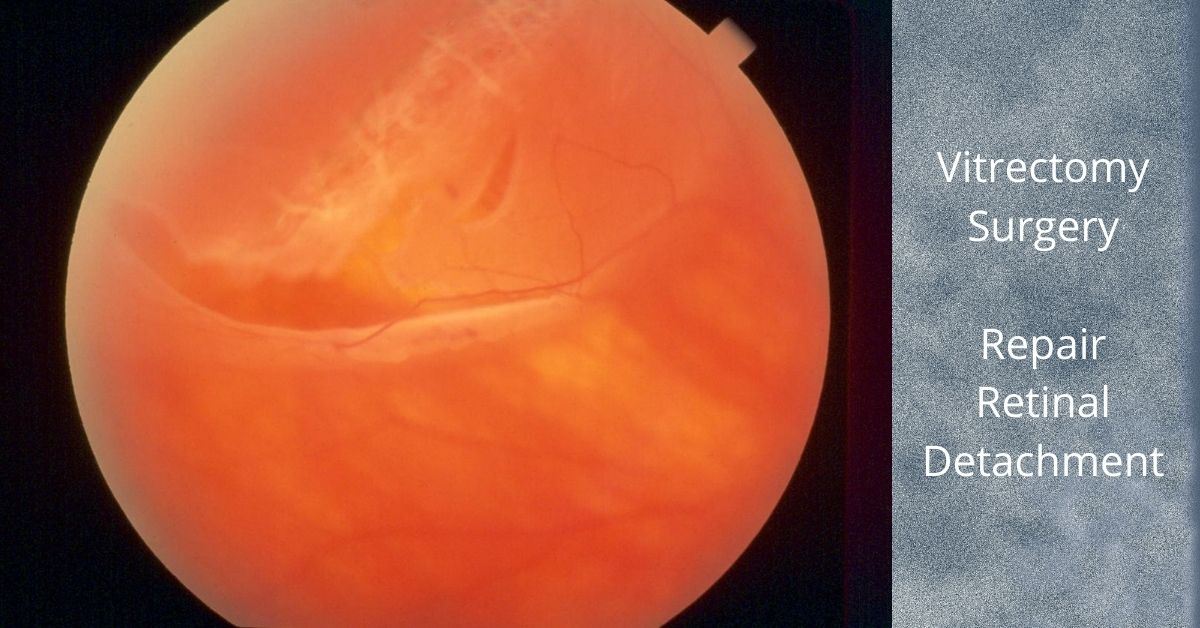
For dense vitreous hemorrhages, it may indeed be prudent to consider a vitrectomy to remove the blood to restore vision, but more importantly, and to allow the retina specialist to perform a thorough examination of the eye with the blood removed.

Retinal Tears and Bleeding

As an example, a retinal tear can cause a vitreous hemorrhage if the tear rips across a retinal blood vessel and causes bleeding.  There may be too much blood to actually visualize a retinal tear.  Keep in mind, retinal tears can lead to retinal detachments.

Retinal detachments can be potentially blinding and, in cases of all retinal tears, we would like to treat the tear before a retinal detachment develops.

Vitreous Hemorrhage Can Resolve

Blood in the vitreous usually absorbs and can clear on with a vitrectomy.  There is no specific timing of this and it can take weeks or even months.  Sometimes, blood does not clear on its own and vitrectomy may be necessary. 

If you would like to schedule an appointment, please call us (877) 245.2020.

Nader Moinfar, M.D., M.P.H.
Retina Specialist
Orlando, FL

Treatment of Diabetic Macular Edema (DME)

Diabetic macular edema is a common complication of diabetic retinopathy where fluid accumulates in the macula and blurs the vision.

Diabetic macular edema (DME) is a condition characterized by the accumulation of fluid in the macula, the central part of the retina responsible for sharp, detailed vision. It is a common complication of diabetic retinopathy, a disease affecting the blood vessels of the retina. The management of diabetic macular edema (DME) involves various treatment approaches aimed at reducing macular edema and preserving vision.

Anti-VEGF Injections – Treatment of Diabetic Macular Edema

One of the primary treatment options for DME is intravitreal injections of anti-vascular endothelial growth factor (anti-VEGF) medications. Drugs like ranibizumab, bevacizumab, and aflibercept are commonly used to inhibit the abnormal growth of blood vessels and reduce leakage, thus decreasing macular edema. These injections are typically administered on a monthly or as-needed basis, with the aim of improving visual acuity and reducing central retinal thickness.

Steroids for Diabetic Macular Edema

Another treatment modality for DME is the use of corticosteroids. Intravitreal injections of triamcinolone acetonide or dexamethasone implants can help suppress inflammation and reduce fluid accumulation in the macula. These steroid injections are usually performed at longer intervals compared to anti-VEGF agents due to their sustained-release properties.

In some cases, laser photocoagulation may be employed to treat DME. Focal laser treatment can be used to seal leaking blood vessels and reduce fluid accumulation. However, this approach is typically reserved for cases where the edema is away from the central macula to avoid damaging central vision.

New Treatments

Recently, a new class of medications called integrin receptor antagonists has shown promise in the treatment of DME. These drugs, such as pegpleranib and abicipar, target specific proteins involved in the pathogenesis of macular edema and are administered via intravitreal injections.

In addition to these specific treatments, managing the underlying diabetes is crucial in preventing and managing DME. Maintaining optimal blood sugar control, blood pressure, and cholesterol levels can help reduce the risk and progression of DME.

Regular eye examinations and early detection of DME are essential for timely intervention and optimal outcomes. Treatment options should be individualized based on the severity of the edema, visual impairment, and patient characteristics. By employing a combination of these treatment modalities and a comprehensive approach to diabetes management, healthcare professionals can improve visual outcomes and enhance the quality of life for individuals living with DME.

If you would like to schedule an appointment, please call us (877) 245.2020.

Nader Moinfar, M.D., M.P.H.
Retina Specialist
Orlando, FL

Dislocated Intraocular Lens

A dislocated intraocular lens is very similar to a dislocated crystalline (natural) lens. The only difference is that the intraocular lens has been inserted during previous cataract surgery.

Cataract surgery is a low-risk surgery and has a success rate of about 95%. But like all surgeries there can be complications, especially if you have other eye or health issues.  

One of those complications is a dislocated intraocular lens (IOL). This is a rare complication and occurs in less than 3% of cases, but it may require additional surgery to repair.

What causes a Dislocated?

Sometimes the dislocation happens shortly after cataract surgery because the zonules, a ring of tiny thread-like fibers that support and hold the lens capsule in place, fail or break. The zonules are made of a connective tissue protein. People who have a connective tissue disorder are more likely to have this complication, and it can also occur in patients who have a history of previous eye surgeries, trauma to the eye, or who take prostate medication.

Other times the IOL dislocation happens gradually over time when the zonules slowly weaken and don’t adequately support the lens capsule and this causes the lens to shift away from the center of the pupil. This happens more often in patients with a history of eye trauma, multiple eye surgeries, especially retinal detachment repair, and complicated original cataract surgery.

Symptoms

The most common symptom of a dislocated IOL is blurry vision. The degree of blurriness depends on how dislocated the IOL is. If the IOL is only slightly dislocated then vision may still be clear, but ghost images or a double image at night may occur when light passes through the edge of the dislocated lens.

Treatments

Treatment depends on the severity of the dislocation. Some dislocations very minor and vision is still good. In these cases, no treatment is necessary.  

If the lens is completely off center and vision is very blurry, then only surgery will correct it. In some cases, the original implanted IOL can be repositioned and secured in place. In other cases when the zonules are extremely weak or damaged, a new IOL designed to be sutured to the wall of the eye (sclera) or to the iris is used.

If the lens has fallen into the vitreous cavity, then a retinal surgeon will need to perform a vitrectomy to remove the vitreous jelly inside the eye and retrieve the dislocated intraocular lens.  

Prognosis

Most patients who have IOL repositioning or IOL replacement have vision of 20/40 or better after surgery. The outcome depends on your health and if you have other eye issues, such as macular degeneration or diabetic retinopathy.  

Recovery for IOL replacement is similar to the recovery time for cataract surgery. The medications are the same and most patients can resume normal activities the day after surgery.

If you would like to schedule an appointment, please call us (877) 245.2020.

Nader Moinfar, M.D., M.P.H.
Retina Specialist
Orlando, FL

Dislocation of the Natural Lens

The fancy term for diplacement of your natural lens in the eye is called ectopia lentis…here’s when it happens.

Your natural lens can become displaced from it’s natural anatomic position within the eye. Intraocular lenses can also become displaced, but this article is limited to dislocation of the natural crystalline lens.

Ectopia lentis is the medical term for the dislocation or displacement of the eye’s natural crystalline lens. The lens may be free-floating in the vitreous or it may be in the anterior chamber or directly on the retina.

This dislocation most commonly occurs after trauma to the eye. It can also be caused by a systemic disease, such as Marfan syndrome. Trauma is the most common cause and most often the result of a direct blow to the eye such as from a baseball or golf ball.

Some rare and inherited genetic diseases can cause ectopia lentis:

  • Weill-Marchesani syndrome
  • Sulfite oxidase deficiency
  • Hyperlysinemia
  • Ehlers-Danlos syndrome
  • Sturge-Weber syndrome
  • Mandibulofacial dysostosis
  • Wildervanck syndrome
  • Conradi syndrome
  • Pfaundler syndrome
  • Crouzon syndrome
  • Pierre Robin syndrome
  • Sprengel deformity

Symptoms of Lens Dislocation

The most significant symptom of ectopia lentis is reduced visual acuity such as poor near or distant vision. The degree of reduction in visual acuity varies with the degree of lens dislocation and the type of dislocation.

If the zonules in the eye are disrupted it can lead to increased curvature of the lens and may result in lenticular myopia or astigmatism. The zonules are tiny thread-like fibers that hold the eye’s lens firmly in place and also tighten the pull the lens to accommodate near vision.

Treatment

Treatment of ectopia lentis depends on where and how far the lens has moved and any resulting complications. In some cases, in which the dislocation is minimal and there is no significant impact on vision observation and close follow-up are the only treatments necessary.

In cases in which vision is affected or there is damage to surrounding structures, surgery may be necessary. The dislocated lens is removed, and an artificial lens is put into place. If there is not enough structural support for the artificial lens then it may need to be sutured to the iris or sclera.

Prognosis

Multiple surgical techniques are available for correction of ectopia lentis and each has its own limitations and associated complications. In some cases, an implanted artificial lens is used, and other patients are treated by removal of the lens and a vitrectomy to remove the vitreous humor and then they use special contact lenses.  

If the lens dislocated due to a genetic disease then the underlying disease must also be treated.

Most patients with ectopia lentis do well and at least 85% achieve a 20/40 or better visual acuity. However, if there is a pre-existing condition such as a corneal disease, glaucoma, diabetic retinopathy, macular degeneration, or a history of retinal detachment, the outcome will not be as favorable.

Close follow-up is essential as is medical management of any complications that may arise. 

If you would like to schedule an appointment, please call us (877) 245.2020.

Nader Moinfar, M.D., M.P.H.
Retina Specialist
Orlando, FL

Vitrectomy for Retinal Detachment Repair

This is the most common way to repair most retinal detachments and has faster healing times.

Vitrectomy for retinal detachment repair is an alternative method to scleral buckle or pneumatic retinopexy.

A vitrectomy is a common surgical repair for a detached retina. The surgery removes the vitreous gel from the eye, pushes the detached retina back into place, and seals any retinal tears in the retina. Then gas is injected into the eye to help keep the retina in place and prevent any fluid from leaking from the repaired tears and into the subretinal space.

The Surgery

A vitrectomy is usually performed using local anesthetic. A heavy sedative is administered and then Lidocaine is injected around the eye to numb it. If patients prefer not to be awake during the procedure they can be given intravenous sedation or a general anesthesia.

The surgery consists of making three small needle-sized incisions (about the width of an eyelash) into the white part of the eye (sclera). Through one incision a constant flow of fluid is passed into the eye. The second incision is used to insert a fiber-optic light to provide illumination of the eye, and the third is for any instruments that are used, including a “vitrector” for the removal of the vitreous.

A vitrector is an oscillating microscopic cutter that slowly removes the vitreous. Once the vitreous is removed, a freezing instrument or a laser is used to seal any tears in the retina.

The vitreous will be replaced with a gas. In time the gas will dissipate and be replaced by the eye’s own fluids (aqueous humor). The vitreous gel does not grow back, but the eye can function with just the fluid that replaces the vitreous. Depending on the type of gas used and its concentration, it can take from three to ten weeks to fully dissipate.

Most retina specialists will perform vitrectomy for retinal detachment repair in an outpatient setting. The surgery time ranges from 20 to 40 minutes, but varies greatly.

Head positioning

Head positioning after surgery is necessary to keep the gas bubble in the proper place. If the retina detached at the bottom, the only way to get the bubble to press against the place of detachment is to place the head down. The bubble walls off the damaged area while it heals and prevents any fluid from flowing through the damaged area of the retina and into the subretinal space.

The gas bubble wiur vision while it is in place, but as it dissipates a line will form across your vision where the newly forming fluid (aqueous humor) is gradually replacing the bubble. The line will move lower each day and the field of vision will get larger.

If you have to position your head down there are pillows, chairs, and mirrors that allow you to see around the room while in a face-down position.

Gas bubble precautions

As long as the gas bubble remains in your eye, you must not fly in an aircraft. The reduced pressure in the cabin will cause the gas bubble to expand. This would be extremely painful and could lead to loss of sight. If you must have general anesthesia for any surgical procedure while the gas bubble is in your eye, you must inform your anesthesiologist so you will not be given nitrous oxide, which would cause a dangerous rise in eye pressure in the eye with the gas bubble.

Safety

Vitrectomy surgery was first developed in 1970 and since then many improvements in instrumentation and technique have dramatically improved the safety of the procedure. Severe complications are rare and the surgical success rate is over 90%.  

Nader Moinfar, M.D., M.P.H.
Retina Specialist
Orlando, FL

If you would like to schedule an appointment, please call us (877) 245.2020.

Cystoid Macular Edema

This type of macular swelling is very common and even occurs after cataract surgery.

What is Cystoid Macular Edema?

Cystoid macular edema, aka CME, affects the functional center of the retina, the macula.  It is another term for macular edema where multiple cyst-like areas of swelling develop.

There are multiple causes of CME to include:

  • Retinal Vascular Occlusions (RVO)
  • Intraocular inflammation (uveitis or iritis)
  • Diabetic retinopathy
  • Uncomplicated cataract surgery

Symptoms of Cystoid Macular Edema

The main symptom of CME is blurry vision.  There may also be some symptoms of distortion, poor color perception and minification, but most patients note blurry vision.

Because this is a condition of the macula, only central vision is affected.  The peripheral or side vision is not involved.

Blurry vision can occur from a variety of eye conditions and is not a specific symptom of cystoid macular edema. 

CME after Cataract Surgery

About 1-3% of patients will develop cystoid macular edema following cataract surgery.  This can occur several weeks following uncomplicated cataract surgery.  Before modern phacoemulsification techniques evolved, CME developed in as many as 10% of patients.

For reasons unknown, fluid starts to accumulate in the macula.  Patients are often frustrated because the vision following surgery was excellent, only to get worse. 

Treatment and Prognosis

CME following cataract surgery is usually self limited, that is, there is full restoration of vision and function after several weeks of treatment.  The condition does not usually recur, but there is increased risk of developing CME in the fellow eye.

Treatment for CME following cataract surgery often involves topical anti-inflammatory eye drops.  Steroid drops are often quite effective.  In more complicated cases, injections of steroid around the eye may be helpful

Because CME can develop from other situations other than cataract surgery, treatments and prognosis vary widely.  For instance, cystoid macular edema following a branch retinal vein occlusion may involve anti-VEGF injections or depot steroids.  

Cystoid macular edema can also be caused by certain glaucoma drops or following complicated cataract surgery.  Surgery may be necessary to treat the macular swelling due to complications of cataract surgery. 

Consultation with a retinal specialist may be needed.

Eye Scan Detects Alzheimer’s Disease

A simple retina scan may soon become a non-invasive method for diagnosing Alzheimer’s disease, and those at risk.

The abnormal proteins that cause Alzheimer’s Disease can build up in the brain two decades before the onset of symptoms and researchers have been searching for ways to detect the disease sooner. Early detection could mean that medications that slow Alzheimer’s progression could be started sooner. The effectiveness of any early treatments could be monitored with eye scans.

Non-Invasive Hi-Res Imaging

Researchers at Duke University and at Washington University School of Medicine in St. Louis have found evidence that Optical Coherence Tomography (OCT) scans can detect the changes in the tiny capillaries and the thinning of the retinal layers in the eye that signal the beginnings of Alzheimer’s disease.

Dr Sharon Fekrat, from Duke University Medical Centre in the US, said: “We’re measuring blood vessels that can’t be seen during a regular eye exam and we’re doing that with relatively new noninvasive technology that takes high-resolution images of very small blood vessels within the retina in just a few minutes.”

Alzheimer’s Causes Retinal Thinning

Microscopic blood vessels form a dense web at the back of the eye inside the retina. In Alzheimer’s patients, researchers saw that that web was less dense, and there was a loss of small retinal blood vessels. The retinal nerve fiber layer was also thinner in patients without Alzheimer’s disease.

Researchers think that reduced levels of acetylcholine in the brain causes some of the symptoms of Alzheimer’s disease. Acetylcholine is one of the neurotransmitter chemicals that nerve cells use to communicate with one another in the brain. Acetylcholinesterase inhibitors prevent the breakdown of acetylcholine and are used to boost the cell-to-cell communication that gets depleted in Alzheimer’s disease.

What is OCT?

Optical coherence tomography is a non-invasive imaging test that uses light waves to take cross-section images of the retina. An OCT shows each of the distinctive layers of the retina. The OCT produces a three-dimensional map of the eye and can show areas of the eye that are abnormal.

The images from an OCT scan are high resolution because they are based on light, rather than sound, as in an ultrasound, or radio frequency, as in an MRI. Because the OCT shows cross-sections of tissues layers, nerve fiber thickness can be measured. 

If you would like to schedule an appointment, please call us (877) 245.2020.

Nader Moinfar, M.D., M.P.H.
Retina Specialist
Orlando, FL

Gas Bubbles and Retina Surgery

Not all retina surgery requires a gas bubble, but here’s when it is necessary for your retina specialist to use a gas.

Not all retina surgery requires injection of a gas bubble.

To safely work on the retina, your retina specialist must be able to safely access the inner layer of the eye. A vitrectomy, the procedure to remove the vitreous, allows the retina specialist specialist to work on the retina. 

A vitrectomy (removal of the vitreous humor gel) is done for the following conditions:

The Gas Bubble

A tamponade in medical terms is something used to close or block a wound or body cavity to stop bleeding or fluid leakage.

Not all retina surgery requires gas to be injected into the eye. The most common use of gas is for repair of a retinal detachment or macular hole. In the case of the retinal detachment (specifically a rhegmatogenous retinal detachment which is caused by a retinal tear or retinal hole), the gas is used to block (tamponade) the migration of fluid to through the tear to underneath the retina.

In the case of a macular hole, the gas is used to allow the hole to slide closed by surface tension.

The gases commonly used are sulfur hexafluoride (SF6) and perfluoropropane (C3F8), and air. Each of the gases dissipates and is replaced by natural fluid, but the time for complete dissipation varies. Air dissipates and is replaced by natural fluid in 5 to 7 days. Sulfur hexafluoride (SF6) dissipates in 10 to 14 days, and perfluoropropane (C3F8), in 55 to 65 days.

The gas bubble blurs your vision while it is in place. As the bubble dissipates you will see a line across your vision where the gas meets the newly forming fluid which is gradually replacing the bubble. The line will move lower each day and your field of vision will get larger as the natural fluid continues to replace the bubble.

As long as any of the gas bubble remains in your eye you must not fly in an airplane because the bubble can expand in the reduced pressure of the cabin causing severe pain and possible loss of sight.

Head Position

Your surgeon will ask you to position yourself in a specific way during healing and that position is dependent on what part of the retina was repaired.

In cases of macular holes, a face down position is common. Head positioning for retinal detachments depends upon the location of the retinal tear(s).

Gas Bubble Injected Last

Most retina surgery is outpatient surgery and can be done under local anesthetic and mild sedation. The sedation is given by IV and is used for anxiety relief and to put you into a relaxed and sleepy state, known as a “twilight state”. In that state you are conscious and still able to hear and follow simple instructions from your surgeon.

General anesthesia can be used for patients with dementia, severe anxiety, or young children.

Most retina surgeries take less than an hour and some less than 30 minutes. The gas bubble is injected as one of the last steps of the surgery.

Recovery

You will have to wear an eye patch for a day or two following surgery. Recovery time depends on the procedure you had, but is generally two to four weeks. An exception is the repair of a complete retinal detachment which could take several months to heal and for vision to stabilize.

If you would like to schedule an appointment, please call us (877) 245.2020.

Nader Moinfar, M.D., M.P.H.
Retina Specialist
Orlando, FL

21st Century Retina Surgery

Most retina surgery is performed on an outpatient basis, and is very similar to cataract surgery.

Retina surgery is complex and requires exacting precision within the microscopic space of the retina.  Recent advances in the size and precision of surgical instruments, microscopic viewing systems, and vitrectomy machines with multiple customizable controls have enabled retina repairs that were impossible just a decade ago.

In addition, these advances in technology have reduced operating times and many retinal repairs take less than an hour and can be done under a local anesthetic and a mild sedative at an outpatient surgical site.

Retina Surgery

To repair retinal holes or tears, remove scar tissue, or reattach a detached retina, the retina specialist must have access to the retina. To do that the vitreous humor gel that fills the eye cavity is removed in a procedure called a vitrectomy. Once the vitreous humor gel is out of the way, the surgeon makes the needed repairs to the retina. 

An EKG and blood pressure, and oxygen sensors are placed to monitor vital signs and sedation is administered through an IV. The sedation puts you in a twilight state in which you are very sleepy and relaxed yet still able to hear your surgeon and respond to simple instructions.

Eye drops are used to numb the eye. Once the eye is numb an eyelid holder is placed to prevent blinking during the surgical procedure. The eyelid holder is not uncomfortable because while your eyes are numb you will not have the sensation of needing to blink.

The Gas Bubble

After your retina is repaired a gas or air bubble is used as a tamponade to prevent the fluid that naturally exudes from inflamed tissues from reaching your retina. The gas bubble walls off and protects the repaired retina while it heals. The gas bubble gradually dissipates and is replaced by natural aqueous fluid.

There are two medical gases that are commonly used. One of them dissipates in 10 to 14 days, whereas the other takes 55 to 65 days to dissipate. If air is used as the bubble, it will absorb within 5 to 7 days.

Depending on your repair, your retina specialist will choose the appropriate medical gas with the correct absorption time.  

The gas bubble makes vision extremely out of focus while it covers the entire vitreous chamber. While it dissipates a line will appear in your vision where the bubble is gradually being replaced by aqueous humor. The line will move further down, and your field of vision will grow larger day by day.

You will be instructed not to fly in an airplane as long as any of the gas bubble remains in your eye. The bubble can expand in the reduced pressure of an airplane cabin causing severe pain and possible loss of sight.

Head Position

You may be asked to keep you head face down during your recovery to keep the gas bubble in the correct position. You can get face down pillows, chairs, and mirrors to help you see things around you while your head is face down. Your insurance might cover the cost of some of the face down recovery equipment.

After Your Retina Surgery

Your eye will be patched after surgery and you will be asked to wear the patch for a day or two following surgery. Recovery time depends on the procedure you had, but ranges from two weeks to several months for a repaired detached retina. 

If you would like to schedule an appointment, please call us (877) 245.2020.

Nader Moinfar, M.D., M.P.H.
Retina Specialist
Orlando, FL

Drug Delivery for Eye Disease

Eye drops are great for only certain eye diseases. There are other routes used to treat your retina.

There are different routes of drug delivery to treat your eye disease.

Not all parts of the eye can be reached by the same route. Eye drops and ointments applied by patients directly to the eye is the route to get to the cornea, conjunctiva, and sclera, but getting to the retina in the back of the eye requires a different route—an injection.

Intravitreal Injections

Only an intravitreal injection will reach the retina because the retina is part of the brain and is protected by the blood-brain barrier.

Intravitreal means that the injection is into the vitreous of the eye. The vitreous is a clear, colorless, jelly-like substance that fills the space between the lens and the retina. Intravitreal injections of anti-VEGF are placed in the vitreous near the retina to treat wet macular degeneration and diabetic retinopathy.

The injections are done in a doctor’s office and the eye is numbed before the injection. They are a safe and effective way to prevent further vision loss.

Eye Drops

Eye drops are the preferred route of drug delivery to treat eye diseases in the front part of the eye. They are commonly used to treat eye infections, allergies, inflammation, glaucoma, and to provide lubrication for dry eyes.

Eye drops come in solutions and suspensions. In a solution the particle sizes are very small and they completely dissolve in the solvent mixture making a solution clear. Suspensions consist of larger particles that are suspended in a solvent. The larger particles will settle to the bottom so suspensions must be shaken before use to re-suspend the therapeutic particles.

If you are applying more than one drop to an eye, allow the first drop to absorb completely before applying a second drop so the medication doesn’t end up running down your cheek.

Medicated Contact Lenses

For some corneal infections a special type of contact lens or a collagen shield is soaked in antibiotics and placed on the eye.

Ointments

Although ointments are greasy and somewhat difficult to apply they are the preferred eye medication for use on infants and very small children because they stay in the eye longer than eye drops. Ointments are also used for nighttime applications of eye medication so the ointment can cover the eye while you sleep.

Sustained Release Drug Systems

These are biodegradable, implanted devices that supply a slow, steady release of a drug over an extended time to maintain the therapeutic level of the drug to the target area. It is new technology and is available for the treatment of glaucoma, vascular occlusions and diabetic retinopathy.

If you would like to schedule an appointment, please call us (877) 245.2020.

Nader Moinfar, M.D., M.P.H.
Retina Specialist
Orlando, FL

Jon Doe