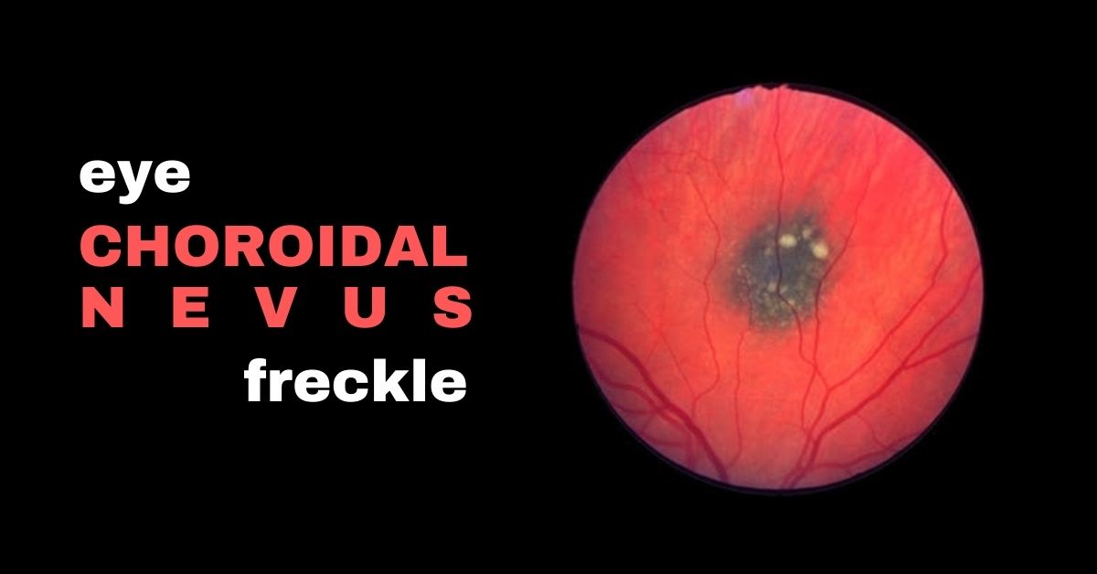
A choroidal nevus is the fancy term for a freckle in the retina.
A nevus is the medical term for mole. Nevi is the plural of nevus. A nevus can appear on your skin, the surface of the eye or inside the eye.
When a nevus is inside the eye it is called a choroidal nevus because those types are found under the retina in the layer of tissue called the choroid.
A nevus is a cluster of melanocytes—the cells that produce melanin. They are the pigment that colors hair, skin, and eyes. They are much more common in people with lighter skin tones.
They are often referred to as eye freckles and the ones that appear inside the eye are discovered during routine eye exams and are usually harmless and most of them won’t affect your vision or cause any problems and they require no treatment except monitoring the nevus for changes.
Three Types of Eye Nevi
- A nevus in the white part of your eye, conjunctival nevus, are common and range from yellow to brown and can lighten or darken over time.
- Nevi in the iris (the colored part of your eye) are tiny, dark brown flecks on the surface of the iris and can grow larger over time but are usually harmless and usually do not become melanomas.
- A choroidal nevus is typically gray, but can be brown, yellow, or variably pigmented. They are also mostly harmless.
Most nevi do not need to be treated and will not affect your vision or lead to any other health problems. The only reason you might need treatment is if your doctor suspects the nevus might be a melanoma.
What Causes a Nevus?
The cause of eye nevi is not known, but there are associations between eye nevi and UV light. Wearing sunglasses to protect your eyes from ultraviolet light is always recommended.
A Choroida Nevus can be Suspicious for Cancer
A very small percentage of choroidal nevi have features that make them at a higher risk of growing into a melanoma. Those choroidal nevi should be monitored by your doctor through regular eye exams. Very rarely, a choroidal nevus may leak fluid or be linked to abnormal blood vessel growth.
There are five factors that signal the choroidal nevus should be closely monitored:
- Thickness of greater than 2.00 mm
- Subretinal fluid
- Visual symptoms
- Orange pigment of the nevus
- A part of the nevus touching the optic nerve
Very often, suspicious cases are referred to a retina specialist or ocular oncologist for further evaluation.
If you would like to schedule an appointment, please call us (877) 245.2020.
