What is the Vitreous?

The vitreous, also known as the vitreous humor, is a gel-like substance that fills the inside of the eye, occupying the space between the lens and the retina. It plays a crucial role in the development of the eye and in supporting the overall structure of the eye after we are born.

Composed mainly of water, the vitreous humor is a transparent, colorless gel that consists of a network of collagen fibers and various other molecules. It is synthesized during the early stages of development and persists throughout life. It is not renewed.

Function

The primary function of the gel is to provide mechanical support to the eyeball. It helps maintain the spherical shape of the eye, which is essential for proper focusing of light onto the retina. Additionally, the vitreous acts as a shock absorber, protecting the delicate structures within the eye from external forces or impact.

Moreover, this clear gel plays a role in the optical system of the eye. By virtue of its transparency, it transmits light, allowing it be focused on the retina. The retina converts light into electrical signals that are then transmitted to the brain, thus creating “vision.” The smooth, uniform consistency of the vitreous helps to ensure that light rays are not scattered or distorted as they travel through the eye.

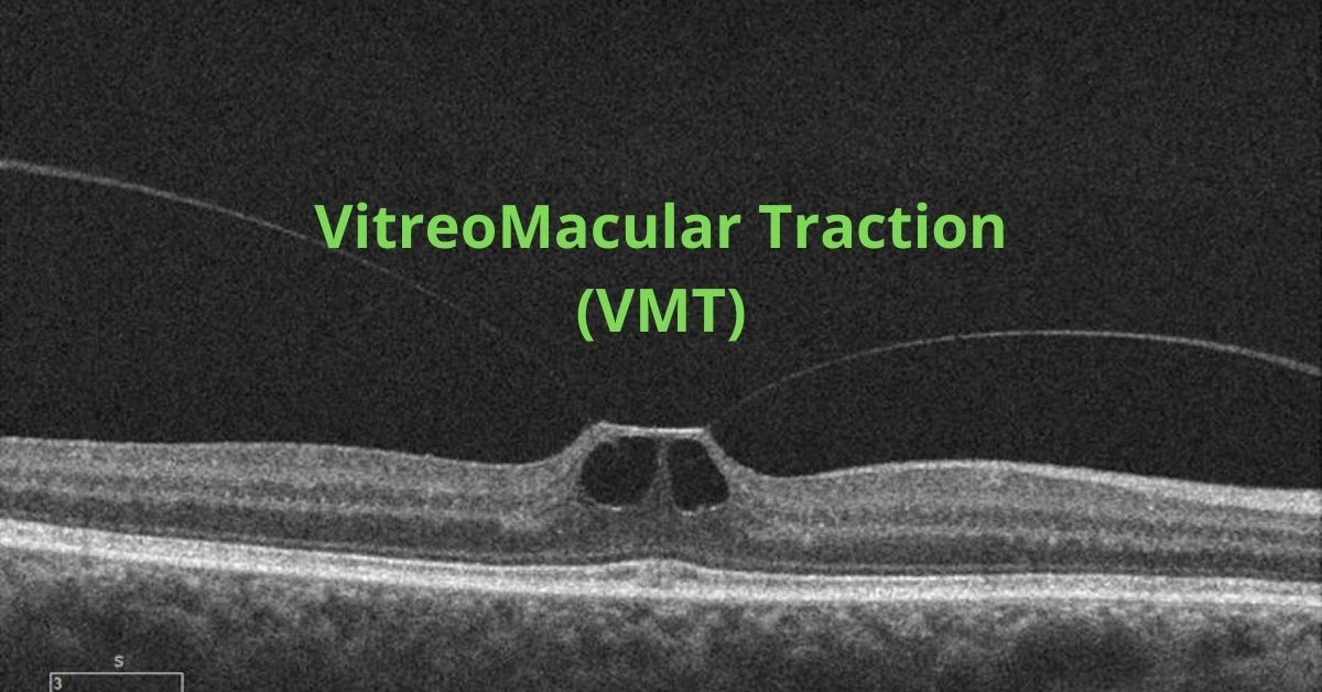
Posterior Vitreous Detachment

While the vitreous is mostly a stable, gel-like substance, it can undergo changes that can impact vision. With age, the vitreous may gradually shrink and develop small pockets of fluid. This is known as a posterior vitreous detachment (PVD). This can lead to the formation of floaters, which are perceived as tiny specks or threads that seem to float across the field of vision. Although floaters are usually harmless, a sudden onset of numerous floaters or flashes of light may be a sign of a retinal tear. A retinal tear can cause a retinal detachment, which requires immediate medical attention. Sudden onset of floaters should always be checked by your eye doctor.

The vitreous is most important for normal development of the eye. Once we are born, it indirectly maintains various functions of the retina and the lens. Understanding the role of the vitreous can help appreciate its importance in maintaining healthy vision and identifying potential eye problems.

If you would like to schedule an appointment, please call us (877) 245.2020.

Nader Moinfar, M.D., M.P.H.
Retina Specialist
Orlando, FL

What is the Retina?

The retina is the light sensitive tissue that translates light into electrical signals for the brain. That’s how we see.

The retina is a highly specialized layer of tissue that lines the inside of the eye. As part of the central nervous system, it serves as a crucial component of the visual system. Its primary function is to convert light into electrical signals that can be translated to “vision” by our brain.

It consists of several layers of cells, each with a specific role in the process of vision. The outermost layer contains the photoreceptor cells, known as rods and cones. Rods are responsible for vision in low-light conditions and are highly sensitive to light, while cones are responsible for color vision and visual acuity in brighter light conditions. Cones are further classified into three types: red, green, and blue cones, allowing us to perceive a wide range of colors.

Converts Light to Electrical Signals

When light enters the eye, it passes through the cornea and lens, which help focus the light onto the retina. The rods and cones in the retina absorb the light and convert it into electrical signals. This process involves the activation of light-sensitive pigments within the photoreceptor cells, triggering a cascade of chemical reactions that generate electrical signals.

The electrical signals produced by the rods and cones are then processed by several layers of cells within the retina. Bipolar cells receive the signals from the photoreceptor cells and transmit them to ganglion cells, which are the final layer of cells in the retina. The ganglion cells collect the signals and their axons form the optic nerve, which carries the visual information from the retina to the brain.

Highly Specialized Cells Sharpen Vision

Within the retina, there are also other types of cells, such as horizontal cells and amacrine cells, which help refine and modulate the visual signals. These cells play a role in enhancing contrast, adjusting sensitivity to light, and facilitating communication between different parts of the retina.

The central region of the retina is known as the macula, which contains a high concentration of cones. At the center of the macula is a small depression called the fovea, where visual acuity is highest. The fovea contains a high density of cones, allowing for detailed and sharp central vision.

For us to see, all layers of the retina must be healthy, maintain normal blood supply and remain attached.

If you would like to schedule an appointment, please call us (877) 245.2020.

Nader Moinfar, M.D., M.P.H.
Retina Specialist
Orlando, FL

What are Eye Floaters?

Vitreous floaters, eye floaters or just floaters can be annoying or a dangerous threat to your vision.

Eye floaters, or vitreous opacities, are common but not always benign. They also affect us all differently, from a mere annoyance to visually impressive.

Floaters are the common name for dark specks or spots which move to and fro in your vision.  They move with eye movement in your field of vision.  They may or may not be associated with clumps or strands of protein floating in your vitreous. These clumps or strands may represent “cob-webs” also seen in your vision.

There are many causes of floaters.

What is the Vitreous?

The vitreous is the substance that fills the vitreous cavity.  The vitreous cavity, or posterior chamber, is the largest compartment of the eye and is located between the lens and the retina.  The retina is the inside lining of the eye. 

Normally, the vitreous is optically clear, color-less and has the consistency of a gel.  The vitreous is composed mostly of water, but has about 5% protein. 

The vitreous is essential for normal embryologic development of the eye.  Once we are born, we are not sure of any true functions of the vitreous although many believe it is essential in oxygenation of the tissues of eye, such as the lens.

The vitreous may absorb energy.  For instance when you are engaged in sports, running, or suffer a head injury, the vitreous may be absorbing shock waves.  This is unproven.

What are floaters?

The vitreous is never regenerated.  While the fluid component does renew (aqueous humor), the proteins do not.  With time and normal aging, the proteins can denature or breakdown and the vitreous becomes more water-like.  

The thinner, more fluid, vitreous means that the vitreous changes from a thicker gel to a more watery substance.  The proteins can more easily coalesce and clump together casting shadows on the retina…more commonly known as floaters. 

Floaters can also be a result of retinal disease, blood or inflammation.  Floaters from any cause are indistinguishable from one another (i.e. you can’t tell the difference between floaters due to blood vs. inflammation).  Only your eye doctor can tell the difference after a thorough dilated examination of your eye.

It is recommended that you alert your eye care professional should you ever experience a sudden increase in floaters.  New floaters could be a sign of a potentially blinding condition. 

Treatment of Floaters

In general, the best treatment for floaters is to treat the underlying disease.  Persistent floaters can be bothersome.  Removal via vitrectomy may be the only solution for clearing the floaters from your vision.

Some doctors may offer Nd:Yag laser for the treatment of floaters.  There are limitations to this type of treatment and not all retina specialists view this treatment equally. 

If you would like to schedule an appointment, please call us (877) 245.2020.

Nader Moinfar, M.D., M.P.H.
Retina Specialist
Orlando, FL

Vitreomacular Traction Syndrome

VMT is very similar to macular pucker and causes the same changes to your vision.

Vitreomacular traction (VMT) is a complication that arises from a normal process. That normal process is posterior vitreous detachment (PVD) and it happens to everyone as they age. By about age 40 or 50 the vitreous gel that fills the eye begins to change. It begins to shrink and lose fluid and strands of the gel can drift through the eye. These strands can be seen as dark strings or spots that float around in your field of view and are called floaters.

The vitreous gel eventually completely separates from the retina. This is perfectly normal and happens to most people by the age of 70, that is, a PVD is a completely normal event.

Problems arise only if the vitreous gel is strongly attached to the retina, specifically at the macula. If that is the case then when the vitreous gel shrinks it can pull on the retina.

That sticking process is called vitreomacular traction and is very similar to an epiretinal membrane.

The pulling and tugging on the center of the retina where the macula is located can damage the macula and cause vision loss if left untreated.

In healthy eyes, VMT is not common. Certain eye conditions or diseases put people at a higher risk of developing VMT. Those conditions and diseases include:

  • High myopia which is extreme nearsightedness
  • Age-related macular degeneration which is a breakdown of the tissues in the back of the eye
  • Diabetic eye disease which affects the blood vessels in the back of the eye
  • Retinal vein occlusion which is a blockage of veins in the retina

Symptoms

The most common symptoms of VMT include:

  • Distorted vision that makes straight lines appear wavy, blurry or have blank spots
  • Seeing lots of flashes of light in your vision
  • Seeing objects as smaller than their actual size

It is important to see an ophthalmologist for an evaluation when you first notice any of these symptoms.

Diagnosis

To diagnose vitreomacular traction (VMT) your ophthalmologist will use tests such as optical coherence tomography (OCT) which uses light waves to take pictures of the various layers of the retina and will show any damage to the macula.

Fluorescein angiography may also be used. This is an imaging test to view how well the blood is circulating inside the retina and to find any macula swelling. It uses a medical imaging dye injected into the arm that circulates inside the retina while a special camera photographs the progress of the dye as it moves through the blood vessels in the eye.

An ultrasound scan may also be used so your ophthalmologist can get a better view of the location of the sticking point between the vitreous and the macula.

Treatment

Some cases do not require treatment and will resolve on their own, but you will be asked to monitor your vision at home with a grid of lines to make sure the VMT does not progress. If the lines on the grid begin to appear wavy or have missing areas then you will most likely require treatment.

Surgery to remove the vitreous and replace it with a saline solution may be needed to prevent macular holes, puckers or macular swelling from developing or worsening.

Some people are candidates for medication treatments. A medication that dissolves the proteins that link the vitreous to the macula can be injected into the eye. Usually only one injection is needed.

Prognosis

Most patients with VMT maintain good visual acuity in the affected eye even if treatment is required.

If you would like to schedule an appointment, please call us (877) 245.2020.

Nader Moinfar, M.D., M.P.H.
Retina Specialist
Orlando, FL

What is a Choroidal Nevus?

A freckle in your is usually benign, but there is a concern about choroidal nevi.

A choroidal nevus is the fancy term for a freckle in the retina.

A nevus is the medical term for mole. Nevi is the plural of nevus. A nevus can appear on your skin, the surface of the eye or inside the eye. 

When a nevus is inside the eye it is called a choroidal nevus because those types are found under the retina in the layer of tissue called the choroid.

A nevus is a cluster of melanocytes—the cells that produce melanin. They are the pigment that colors hair, skin, and eyes. They are much more common in people with lighter skin tones.

They are often referred to as eye freckles and the ones that appear inside the eye are discovered during routine eye exams and are usually harmless and most of them won’t affect your vision or cause any problems and they require no treatment except monitoring the nevus for changes.

Three Types of Eye Nevi

  • A nevus in the white part of your eye, conjunctival nevus, are common and range from yellow to brown and can lighten or darken over time. 
  • Nevi in the iris (the colored part of your eye) are tiny, dark brown flecks on the surface of the iris and can grow larger over time but are usually harmless and usually do not become melanomas. 
  • A choroidal nevus is typically gray, but can be brown, yellow, or variably pigmented. They are also mostly harmless.  

Most nevi do not need to be treated and will not affect your vision or lead to any other health problems. The only reason you might need treatment is if your doctor suspects the nevus might be a melanoma.

What Causes a Nevus?

The cause of eye nevi is not known, but there are associations between eye nevi and UV light. Wearing sunglasses to protect your eyes from ultraviolet light is always recommended.

A Choroida Nevus can be Suspicious for Cancer

A very small percentage of choroidal nevi have features that make them at a higher risk of growing into a melanoma. Those choroidal nevi should be monitored by your doctor through regular eye exams. Very rarely, a choroidal nevus may leak fluid or be linked to abnormal blood vessel growth.

There are five factors that signal the choroidal nevus should be closely monitored:

  • Thickness of greater than 2.00 mm
  • Subretinal fluid
  • Visual symptoms
  • Orange pigment of the nevus
  • A part of the nevus touching the optic nerve

Very often, suspicious cases are referred to a retina specialist or ocular oncologist for further evaluation.

If you would like to schedule an appointment, please call us (877) 245.2020.

Nader Moinfar, M.D., M.P.H.
Retina Specialist
Orlando, FL

Should You Be Taking Vitamins for AMD?

Are these vitamins for you? Here’s who benefits the most and who shoujld be taking them.

Do you take vitamins for AMD?

The National Eye Institute conducted the Age-Related Eye Disease Study (AREDS) and AREDS2 to study both cataract and age-related macular degeneration (AMD). The study followed participants for five years and tested whether taking nutritional supplements (vitamins for AMD) could prevent or slow these two eye diseases.

AREDS Results

At the end of the five-year study, the scientists found that people at high risk for developing advanced AMD—those with intermediate AMD, and those with advanced AMD in one eye only—reduced their risk of developing advanced by about 25 percent when treated with the combination of antioxidants and zinc + copper. 

The AREDS formula also reduced the risk of central vision loss by 19% in the same group. None of the formulations decreased cataract risk.

Who Benefits the Most?

The study found that those who benefit most from taking vitamins for AMD are people at high risk for developing macular degeneration and who meet these two conditions:

  • Intermediate AMD in one or both eyes
  • Advanced AMD in only one eye

AREDS Vitamins for AMD

The AREDS 2 vitamins do not contain beta carotene because in the study researchers found that beta carotene in high amounts was associated with an increased risk of lung cancer in current and former smokers.

You can purchase AREDS 2 vitamins or take the supplements separately. The following is the AREDS 2 formulation:

  •  Vitamin C (ascorbic acid) 500 mg
  • Vitamin E 400 international units (IU)
  • Lutein 10 mg
  • Zeaxanthin 2 mg
  • Zinc (as zinc oxide) 80 mg 
  • Copper (as cupric oxide) 2 mg

Healthy Foods

Taking vitamins is just one step in creating a healthy environment for your eyes. Eating a healthy diet is an essential second step. Include lots of dark leafy greens, like spinach and kale, along with other colorful vegetables and fruits. Colorful vegetables and fruits provide a wide range of phytonutrients along with carotenoids that your body converts into vitamin A.  

Smoking puts you at a higher risk for AMD, so if you smoke, then stopping will be another step toward eye health.

Consult Your Doctor

Talk with your eye doctor about your primary care physician about how taking the AREDS or AREDS 2 vitamins will affect you.  

Nader Moinfar, M.D., M.P.H.
Retina Specialist
Orlando, FL

If you would like to schedule an appointment, please call us (877) 245.2020.

Saving Your Vision: Diabetic Retinopathy

November is National Diabetes Awareness month.

If you have diabetes, take a few minutes to learn about diabetic retinopathy and how you can take steps to help keep your eyes healthy.

Diabetic retinopathy is the most common cause of vision impairment and blindness among working-age adults in the U.S. It occurs when high blood glucose levels damage the blood vessels at the back of the eye causing them to become weak and leaky and damage the retina.

If not found and treated early, diabetic retinopathy can cause permanent vision loss. That’s why it is important to manage your diabetes and stay alert to the early signs and symptoms of diabetic retinopathy.

Types of Diabetic Retinopathy

There are two main types of diabetic retinopathy—NPDR (non-proliferative diabetic retinopathy) and PDR (proliferative diabetic retinopathy). The “proliferative” refers to new blood vessel growth, so in non-proliferative, there is not yet new blood vessel growth.   

NPDR is the early stage of diabetic retinopathy. At this stage, high glucose levels have weakened the blood vessels and, in time, may cause bulges in the weakened vessels (microaneurysms) that may leak fluid into the retina and the leakage may swell the macula or some blood vessels may begin to close. So this is an ongoing disease cycle that can be slowed or stopped if caught early.

PDR is the more advanced stage of diabetic retinopathy. The NPDR has weakened the blood vessels or closed them, so new blood vessels have grown in an attempt to supply oxygen to the retina, but these new vessels are fragile, and they leak and damage the retina. 

PDR can lead to vitreous hemorrhage, traction retinal detachment, or neovascular glaucoma.

Stay Alert

Anyone with type 1 or type 2 diabetes, or women who have gestational diabetes during pregnancy, can develop diabetic retinopathy. The risk increases when blood sugar, blood pressure, and cholesterol levels are not under control.

In the early stages diabetic retinopathy may not have symptoms, so it’s important to have a comprehensive dilated eye exam once a year. That way diabetic retinopathy can be diagnosed and treated before symptoms appear.   

Symptoms that indicate diabetic retinopathy has progressed are:

  • Blurry vision
  • Vision that goes from blurry to clear
  • Increased number of floaters
  • Poor night vision
  • Spots or blank areas in your central vision
  • Colors appear faded

Diabetic Retinopathy Treatment

The best treatment happens before you develop symptoms. Strict control of your blood sugar will significantly reduce your risk of vision loss from diabetic retinopathy. If you have high blood pressure and kidney problems, make sure those are kept under control too.

Injections in the eye of anti-VEGF medication can stop the growth of new blood vessels. VEGF stands for vascular endothelial growth factor, and it is a signal protein your body produces that stimulates new blood vessel formation, but the new vessels are weak and leaking vessels that damage your eyes, and anti-VEGF injections will stop that abnormal blood vessel growth.

Laser surgery and vitrectomy are other treatments for advanced PDR. But the most important thing to remember if you have diabetes, is that control of your blood sugar and early diagnosis and treatment of diabetic retinopathy are the best protections against vision loss.

If you would like to schedule an appointment, please call us (877) 245.2020.

Nader Moinfar, M.D., M.P.H.
Retina Specialist
Orlando, FL

Eating Carrots to Improve Your Eye Health

Do carrots really help your vision? Here’s how carrots improve your eye health.

Getting enough vitamin A is essential to maintain eye health and carrots are an excellent natural source of vitamin A, but eating a diet overloaded with carrots or other sources of vitamin A won’t improve your eyesight.

Eye health and good eyesight are different things. Healthy eyes can still need glasses to clearly focus on near or far objects because the shape of the cornea or the eyeball causes nearsightedness, farsightedness, or astigmatism.  No amount of vitamin A will change that.

Vitamin A and Eye Health

Vitamin A is converted into rhodopsin, the photopigment in the rods of your retina that enable vision in low-light. In humans and many animals rhodopsin is required for vision in dim light. 

Rhodopsin converts light into electrical nerve signals within the photoreceptor rods and those electrical signals are sent to the brain via the optic nerve. 

Vitamin A also supports the healthy functioning of the eye’s conjunctival membranes and the corneas.

Vitamin A deficiencies are rare in the U.S. and other developed countries. Most diets in developed countries contain adequate amounts of vitamin A and vitamin A can be stored by the body in the liver. 

Excellent Sources of Vitamin A

Sweet potatoes, spinach, pumpkin, carrots, milk, cheese, cantaloupe, mangos, eggs, and sweet red peppers are all excellent sources of vitamin A.

Vitamins and Oxidative Stress

The human body produces oxygen-derived free radicals as part of the metabolic process, part of breathing and burning energy, and free radicals are essential to life, but their number must be balanced out with antioxidants for optimal health.

A diet rich in antioxidants such as vitamins A, E, and carotenoids such as beta carotene, lycopene, and lutein will help your body scavenge free radicals, terminating their chain reactions before they cause cellular damage.

There is a peptide (glutathione) and some antioxidant enzymes produced by the body specifically to scavenge free radicals, but consuming a diet of antioxidant-rich fruits and vegetables, will also be a diet low in saturated fat and cholesterol, so eating lots of high-antioxidant fruits and vegetables will not only add to your body’s antioxidant levels, but give your overall health a boost.  

If you would like to schedule an appointment, please call us (877) 245.2020.

Nader Moinfar, M.D., M.P.H.
Retina Specialist
Orlando, FL

Jon Doe