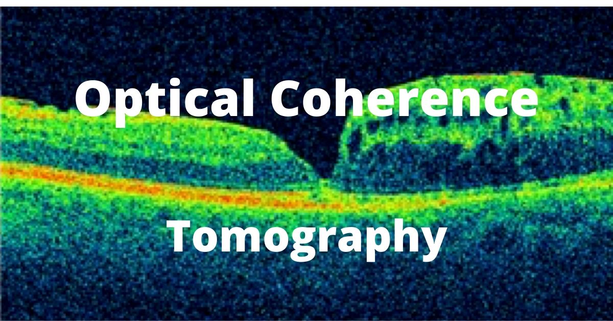
OCT, or optical coherence tomography, is another diagnostic test used by a retina specialist. A fluorescein angiogram has been discussed previously and it also provides valuable information about the health of the retina.
An OCT uses light waves to study the different layers of the retina and also provides information about the surface of the retina.
Topography is the study of the surface (in this case the retina) where tomography allows study of a tissue in cross-section.
OCT Studies Retinal Diseases
Common uses for an optical coherence tomogram (OCT) include:
Macular Degeneration
Macular Holes
Macular Pucker
Macular Edema
Diabetic Retinopathy
Retinal Vascular Occlusions (RVO)
The OCT allows the retina specialist to diagnose and problem, but also allows me to evaluate progression of a disease and monitor if a treatment is effective.
For instance, after intra-vitreal injections of VEGF, an OCT allows me to determine if the treatment is reducing the retinal swelling caused by either diabetes or macular degeneration.
Other Uses of OCT
Retinal specialists are not the only ophthalmologists who use this state of the art technology. Using OCT technology to examine the optic nerve is a great way to diagnose and monitor progression of glaucoma.
Performing the Optical Coherence Tomography
The test is painless and non-invasive. No dye or contrast is used. There is no injection (unlike a fluorescein angiogram).
An OCT requires that you are able to sit and place your chin of the machine while keeping your eyes and head very still. A target is provided to keep the eyes still. Nothing will touch your eye.
The whole process takes a few minutes per eye. Usually we prefer the eyes to be dilated. On occasion, very dense cataracts or vitreous hemorrhage (bleeding in the vitreous) prevents a good test result. Remember the test relies on light rays entering your eye. Both dense cataracts and vitreous hemorrhage can block light.
If you would like to schedule an appointment, please call us (877) 245.2020.
