What is Legal Blindness?

Legal blindness can be caused by a variety of diseases. Here’s how it is defined and links to resources.

Legal blindness is a legal definition that is used to refer to patients with vision loss who qualify for government assistance. This is not total blindness.

Legal blindness is defined as a patient’s best visual acuity (aka best corrected) in either eye is 20/200 or less or the size of the visual field (peripheral vision) is less than 20 degrees. In other words, if both eyes do not see well, the vision is no better than 20/200 in better seeing eye.

“Best corrected” refers to the use of the best corrective lens/contact lens for the patient.

A central visual acuity of 20/200 or worse means that a legally blind person must be 20 feet away from an object to see what a “normal” person can see from 200 feet away.

Loss of peripheral vision can also qualify as legal blindness. Most patients normal peripheral vision can see at least 140 degrees without turning the head. Legally blind peripheral vision is less than 20 degrees.

Causes of Legal Blindness

  • Age-related macular degeneration (ARMD) is one of the leading causes of legal blindness in Americans aged 60 and older. ARMD affects the macula (functional center of the retina) and therefore decreases central vision.
  • Cataracts will affect almost everyone by the age of 80. Cataracts blur both central and peripheral vision (though reversible with cataract surgery).
  • Diabetic retinopathy is another leading cause of blindness and affects the blood vessels in the back of the retina. Diabetic retinopathy causes either blurring of the central vision (most common), but can affect peripheral vision, too.
  • Glaucoma is a progressive disease that damages the optic nerve. Glaucoma primarily causes visual field loss and, only in the late stages, affects central vision.

NOTE: All of the disease described above have available treatments which, in most cases, can preserve vision or slow down progression if diagnosed timely.

Low Vision Aids

May low-vision aids and devices are available to assist individuals who are legally blind. Both near and distance vision can be improved with low-vision aids.

For example, desktop, stand-alone and hand-held magnifiers are available. These may be helpful for close range work such as reading or computer use. Prices vary up to several hundred dollars.

Wearable devices that magnify are also available. Mounted binoculars, wearable HD autofocus cameras with TV viewing and electronic headsets with built-in cameras can help patients with central and peripheral vision loss. Prices vary up to several thousand dollars.

Resources

There are many governmental and non-governmental resources for those who are legally blind.

For example, the American Foundation for the Blind (AFB) can assist those with low-vision. Founded in 1921, the AFB ensures that patients who are blind, legally blind or otherwise visually impaired have access to educational materails, technology and legal information.

Here are additional resources that help blind, legal blindness and visually impaired.

Low Vision Evaluation

If you feel you suffer from legal blindness or need more information, a low-vision examination is the first place to start. Eye doctors specializing in low vision can advise and educate you about the best low-vision aids for your specific visual needs.

If you would like to schedule an appointment, please call us (877) 245.2020.

Nader Moinfar, M.D., M.P.H.
Retina Specialist
Orlando, FL

Possible COVID-19 Treatment can Cause Eye Damage

Hydroxychloroquine is FDA approved for certain autoimmune diseases and malaria, but has the potential for toxicity…

Hydroxychloroquine has long been used to safely treat patients with malaria and certain autoimmune diseases such as lupus erythematosus and rheumatoid arthritis, but NOT COVID019.

In the news, there have been small, inconclusive studies using two malaria and autoimmune medications to treat COVID-19 (coronavirus 2019). Some studies suggest the medications might be helpful, while other studies saw no difference.

Currently, there is not enough medical data to prove that hydroxychloroquine and chloroquine work for COVID-19.

Hydroxychloroquine Approved for Malaria

Chloroquine was approved by the FDA in 1949 as a treatment for malaria, an infection caused by a parasite. Hydroxychloroquine was approved by the FDA in 1955 for the treatment of malaria. Hydroxychloroquine is preferred over chloroquine because it has fewer side effects.

Both drugs are available in pill form. Plaquenil is also approved for long-term use in rheumatoid arthritis and lupus.

The side effects of both medications include:

  • Retinal damage and irreversible vision loss (Plaquenil Toxicity)
  • Headache
  • Abnormal heart rhythm
  • Muscle weakness or nerve pain
  • Gastrointestinal issues (nausea, cramping, vomiting, and diarrhea)
  • Hypoglycemia (low blood glucose)
  • Worsening of psoriasis

Retinal Damage | Plaquenil Toxicity

The overall prevalence of chloroquine/hydroxychloroquine retinopathy is 7.5%. Retinal damage is one of the most serious adverse effects of hydroxychloroquine (HCQ) since the retinal damage caused by the drug is irreversible. Chloroquine/hydroxychloroquine retinopathy is more common with higher daily doses and long-term use.

A baseline retinal examination is recommended for all patients taking HCQ, along with regular monitoring examinations. The baseline and monitoring examinations should include the following:

  • Uncorrected visual acuity test
  • Best-corrected visual acuity test
  • Examination with ophthalmoscope
  • Examination using slit-lamp
  • Optical Coherence Tomography

The FDA previously issued an Emergency Use Authorization (EUA) that allows physicians to request a supply of hydroxychloroquine or chloroquine for hospitalized patients with COVID-19 who are unable to join a clinical trial. 

As of this writing 4/24/20, the FDA now warns against the use of hydroxychloroquine for the treatment of COVID-19 after reports of irregular heart rhythms and deaths.

If you are presently taking Plaquenil, talk to your prescribing doctor to insure that you are getting regular eye exams.

If you would like to schedule an appointment, please call us (877) 245.2020.

Nader Moinfar, M.D., M.P.H.
Retina Specialist
Orlando, FL

Elective Surgery May Stop

Here’s why your follow up visit or even your surgery may be canceled.

As the COVID-19 pandemic continues to spread, the American Academy of Ophthalmology (AAO), the Centers for Disease Control and Prevention (CDC) and the American Hospital Association have issued recommendations aimed at reducing exposure and preserving healthcare resources, aka, “flattening the curve.”

Will this affect all eye doctors?  Will this affect you?

Stop Routine Care

The American Academy of Ophthalmology is recommending that eye doctors stop routine eye care at this time and focus only on those patients with urgent and emergent conditions.  Examples of “routine” care might include glasses prescriptions, routine follow-up or cataract evaluations. 

Please check with your own doctor if you need to be seen.

As a retina specialist, I may not necessarily “need” to see a patient I see every 6 months with diabetes who is not noticing any changes since the last visit, but I may need to evaluate someone who recently lost vision or developed pain.  

By limiting the number of patients who come through our offices, we are protecting our patients, our staff and ourselves from exposure to the virus.  It is our way of increasing the likelihood that a patient may remain in place and limit the spread of this disease. 

Stop Elective Surgery

Both the American Hospital Association and the Centers for Disease Control and Prevention are recommending that hospitals stop performing elective surgery and non-urgent surgical procedures.  Examples of each might include joint replacements and cataract surgery. Non-urgent surgical procedures might include a colonoscopy.

What’s the rationale?  It saves resources.  

Elective surgeries and procedures take up hospital resources.   By canceling or postponing these procedures, for example, hospital beds and staff can be freed to help with more critical patients elsewhere in the hospital system.

Nurses and aides who normally work in an elective surgical center may be needed elsewhere such as the emergency room.

This practice will also limit exposure to healthy patients.  

Don’t forget that while this pandemic is progressing, patients will still need emergency surgery, chemotherapy, and require care for heart attacks and strokes. 

This is an effort to avoid overburdening our hospital resources and “flatten the curve.”

If you would like to schedule an appointment, please call us (877) 245.2020.

Nader Moinfar, M.D., M.P.H.
Retina Specialist
Orlando, FL

Retinal Disease Urgencies & Emergencies

As the pandemic looms, do you know what urgent and emergent eye conditions might mean?

Eye doctors will likely be asked to see only patients with urgent and emergent conditions. Retinal diseases often require emergent or urgent treatment.

Suggestions have been made to limit office visits to those patients with only urgent and emergency conditions.  In addition, hospitals have been canceling elective surgery and procedures to a later date. Both measures are intended to protect healthy individuals from unnecessary exposure to the virus and to preserve healthcare resources.

Retinal specialists treat “urgent” and “emergent” on a daily basis.  Here’s a short list of what a retina specialist treats on a daily basis.  

Disclaimer:  This article reflects my opinion only.  Your own doctor may have a different opinion.  Please check with your doctor if you have questions or if you are having problems with your vision.  In my opinion, any condition which causes a change in vision or pain should warrant an urgent visit to your doctor….call your doctor.  

N. Moinfar, M.D.

Diabetic Retinopathy

Diabetic retinopathy is the disease diabetes causes in the eye.  Most patients will develop some degree of diabetic retinopathy in their lifetime, and because of this, patients need to be seen regularly whether there are changes in their vision or not.

If there are no acute changes in vision since last visit, then there is probably no urgency to get an examination, though this may not be true in patients with proliferative diabetic retinopathy.  You may call your doctor and discuss how long you can wait after the pandemic is over. 

Diabetes can cause sudden loss of vision due to a vitreous hemorrhage.  Your doctor may want to see you quickly.

New Onset Floaters

Usually new onset floaters are due to a posterior vitreous detachment (PVD).  A PVD can cause a retinal tear which can be diagnosed only by prompt examination and, therefore, new onset floaters should be deemed urgent.  Retinal tears can cause a retinal detachment, hence, the concern.

Macular Degeneration

Patients with macular degeneration can develop loss of central vision and/or distortion.  Many patients self monitor at home with the aid of an Amsler grid, a simple way to look for new distortion in their vision.  If you notice new loss of vision and/or worsening distortion, especially if you are receiving anit-VEGF injections, I would recommend you call your doctor for an urgent evaluation.

Complications from Cataract Surgery

Complications from cataract surgery are usually seen by a retina specialist within a few days of occurrence.  Since cataract surgery is deemed elective, there should not be any complications during the pandemic as cataract surgery is being rescheduled to preserve hospital resources and decrease exposure of healthy people to the virus. 

Retinal Detachment

Retinal detachments can be an emergency, especially if there is progressive loss of vision, but the central vision is spared.  The goal of retinal detachment surgery is to diagnose and perform surgery before the central vision is affected.  

If you would like to schedule an appointment, please call us (877) 245.2020.

Nader Moinfar, M.D., M.P.H.
Retina Specialist
Orlando, FL

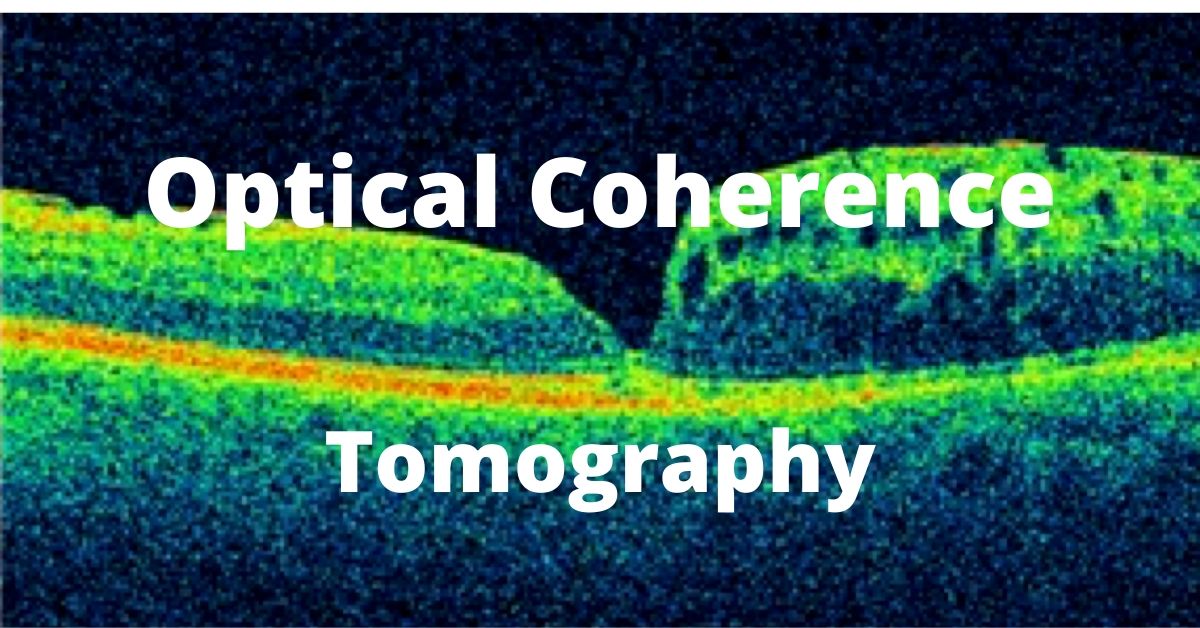
Optical Coherence Tomography (OCT)

OCT, or optical coherence tomography, is another diagnostic test used by a retina specialist. A fluorescein angiogram has been discussed previously and it also provides valuable information about the health of the retina.

An OCT uses light waves to study the different layers of the retina and also provides information about the surface of the retina.

Topography is the study of the surface (in this case the retina) where tomography allows study of a tissue in cross-section.

OCT Studies Retinal Diseases

Common uses for an optical coherence tomogram (OCT) include:

Macular Degeneration
Macular Holes
Macular Pucker
Macular Edema
Diabetic Retinopathy
Retinal Vascular Occlusions (RVO)

The OCT allows the retina specialist to diagnose and problem, but also allows me to evaluate progression of a disease and monitor if a treatment is effective.

For instance, after intra-vitreal injections of VEGF, an OCT allows me to determine if the treatment is reducing the retinal swelling caused by either diabetes or macular degeneration.

Other Uses of OCT

Retinal specialists are not the only ophthalmologists who use this state of the art technology. Using OCT technology to examine the optic nerve is a great way to diagnose and monitor progression of glaucoma.

Performing the Optical Coherence Tomography

The test is painless and non-invasive. No dye or contrast is used. There is no injection (unlike a fluorescein angiogram).

An OCT requires that you are able to sit and place your chin of the machine while keeping your eyes and head very still. A target is provided to keep the eyes still. Nothing will touch your eye.

The whole process takes a few minutes per eye. Usually we prefer the eyes to be dilated. On occasion, very dense cataracts or vitreous hemorrhage (bleeding in the vitreous) prevents a good test result. Remember the test relies on light rays entering your eye. Both dense cataracts and vitreous hemorrhage can block light.

If you would like to schedule an appointment, please call us (877) 245.2020.
Nader Moinfar, M.D., M.P.H.
Retina Specialist
Orlando, FL

What is a Fluorescein Angiogram

Fluorescein angiography is a common diagnostic test used primarily by a retina specialist for certain retinal diseases, such as…

A fluorescein angiogram is a diagnostic test used to study diseases of the retina. While any ophthalmologist may perform this test, a retina specialist uses this single test more than any other type of doctor.

Patients with the following diseases are likely to require a fluorescein angiogram (FA):

Diabetes
ARMD – macular degeneration
Macular edema
Retinal vascular occlusions

As the name implies, fluorescein dye used to study abnormalities in the retina and of its blood supply

The Fluorescein Angiogram

An FA is performed by injecting this vegetable-based dye into a vein. We usually choose an arm vein because it allows easy access for the doctor or technician.

The dye is slowly injected and will reach the eye in about 15 seconds under normal conditions. Patients with heart conditions or abnormalities in blood flow to the eye may experience longer times for the dye to reach the eye. Pictures are taken as the dye flows through the retina.

Abnormalities of retinal blood flow, degeneration of the layers of the retina and retinal swelling can be easily detected with this test.

Is FA Safe?

As with any injectable, there are possible complications. Overall, the fluorescein angiogram is a safe test, but your doctor should talk to about a few possible side effects.

Nausea and vomiting are the most common side effects, but usually occur during the test and pass quickly. Itching and hives can occur and, very rarely, anaphylaxis is possible.

Be aware that the fluorescein dye is unique to ophthalmology and has no relation to diagnostic contrast dyes used in radiologic testing such as MRIs.

If you would like to schedule an appointment, please call us (877) 245.2020.

Nader Moinfar, M.D., M.P.H.
Retina Specialist
Orlando, FL

Dilated Eye Exams

Why you need to have your eyes dilated by a retina specialist.

In order to properly examine your retina, a dilated eye exam is necessary each and every time you come to the office. A dilated eye exam requires use of dilating drops to dilate and enlarge your pupils to allow us to properly diagnose and treat diseases of the retina.

A dilated eye exam is the only way the retina can be examined by direct visualization using a combination of lenses and specialized ophthalmic instruments.

Other methods such as photography, fluorescein angiography, optical coherence tomography (OCT) and ultrasound can be used in conjunction to direct visualization to diagnose and to treat retinal diseases.

As a retina specialist, virtually everyone of my patients needs a dilated eye exam every time they come to the office.

Dilating Eye Drops

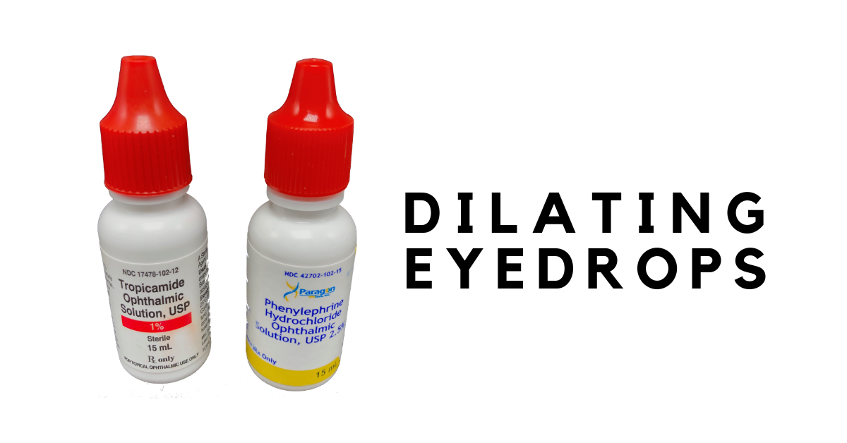
There are several drops used routinely to dilate the eyes. All are marked with a bright red cap to distinguish these eye drops from other types.

In most cases, we only need to use phenylephrine and tropicamide do satisfactorily dilate the pupils. These drops work differently to achieve dilation.

Tropicamide inhibits the small muscle located at the margin of the pupil. The function of the pupillary sphincter muscle is to constrict the iris in bright light and makes the pupil smaller through a purse-string mechanism. Tropicamide prevents this constriction.

Phenylephrine stimulates another set of muscles which function to enlarge the pupil. The iris dilator muscle runs radially along the iris and, when stimulated, pulls the iris open.

Cyclopentolate and atropine can also dilate the pupils but their effects last much longer (days to weeks) and are generally not used for diagnostic purposes.

How Long does Dilation Last?

In most circumstances, your eyes will stay dilated for several hours after the exam. Several factors influence the length of time your eyes will stay dilated, such as:

  • Color of your eyes
  • Your age
  • Type, strength and amount of the medicine
  • Previous eye surgery

Lightly colored eyes, such as blue and hazel, dilate very quickly and stay dilated longer compared to brown eyes.

Brown eyes actually contain brown colored pigment which absorbs the dilating medicine, whereas blue eyes actually contain no pigment and the eyes dilate faster.

Younger eyes tend to take a longer time to dilate. Certain diseases (e.g. iritis) can make dilation take a very long time.

Dilation usually takes at least 20 minutes at a minimum and will reverse after a few hours.

If you would like to schedule an appointment, please call us (877) 245.2020.
Nader Moinfar, M.D., M.P.H.
Retina Specialist
Orlando, FL

Jon Doe