What is FOV?

FOV stands for Floater Only Vitrectomy.  In my experience, this is the common name of the basic operation performed by a retina specialist called vitrectomy.

Vitrectomy and FOV, therefore, are synonyms.  Vitrectomy is the standard operation performed by all retina specialists (for all reasons) whereas FOV refers to a vitrectomy performed for the removal of only (just) floaters.

Vitrectomy

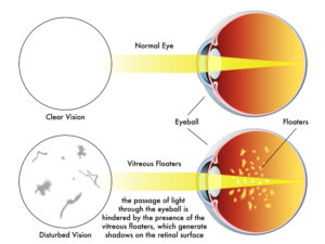
Vitrectomy is the operation to remove the vitreous.  The vitreous is the gel-like substance that fills the back of the eye.  

The vitreous may be removed;

  • To remove a vitreous hemorrhage (blood)
  • Remove a foreign body
  • Repair
    • Retinal detachment
    • Macular hole
    • Epiretinal membrane
  • Remove floaters

Types of FOV or Vitrectomy

All modern types of FOV are basically the same.  They involve the creation of 3 holes in the eye:

  1. Left hand
  2. Right hand
  3. Infusion port to keep the eye filled with saline during the operation

There are slight changes in nomenclature to distinguish the size or thickness of the instruments used to enter the eye.  The size of the sclerotomies (holes) the instruments create naturally vary in size.  The thicker instruments create large holes and vice versa.  20 gauge instruments are the thickest and require sutures to close the sclerotomies.  23 and 25 gauge instrumentation create tinier sclerotomies and suture closure is up to the discretion of the retina specialist.

What Replaces the Vitreous?

The vitreous is cut away rapidly in very tiny miniscule pieces and is replaced by artificial saline solution.  As the operation is proceeding, a tube is constantly infusing saline into the eye.  After the operation is complete, your eye replaces the saline with aqueous humor in a matter of a couple days.

How is FOV Different than Vitrectomy

It’s not.  It’s exactly the same.  After the vitreous is removed, the operation is essentially over.  FOV for floaters is exactly the same as FOV for vitreous hemorrhage – the goal of both is to simply clear opacities from the vitreous to create a clear path for vision.

If you would like to schedule an appointment, please call us (877) 245.2020.

Nader Moinfar, M.D., M.P.H.
Retina Specialist
Orlando, FL

What are Eye Floaters?

Vitreous floaters, eye floaters or just floaters can be annoying or a dangerous threat to your vision.

Eye floaters, or vitreous opacities, are common but not always benign. They also affect us all differently, from a mere annoyance to visually impressive.

Floaters are the common name for dark specks or spots which move to and fro in your vision.  They move with eye movement in your field of vision.  They may or may not be associated with clumps or strands of protein floating in your vitreous. These clumps or strands may represent “cob-webs” also seen in your vision.

There are many causes of floaters.

What is the Vitreous?

The vitreous is the substance that fills the vitreous cavity.  The vitreous cavity, or posterior chamber, is the largest compartment of the eye and is located between the lens and the retina.  The retina is the inside lining of the eye. 

Normally, the vitreous is optically clear, color-less and has the consistency of a gel.  The vitreous is composed mostly of water, but has about 5% protein. 

The vitreous is essential for normal embryologic development of the eye.  Once we are born, we are not sure of any true functions of the vitreous although many believe it is essential in oxygenation of the tissues of eye, such as the lens.

The vitreous may absorb energy.  For instance when you are engaged in sports, running, or suffer a head injury, the vitreous may be absorbing shock waves.  This is unproven.

What are floaters?

The vitreous is never regenerated.  While the fluid component does renew (aqueous humor), the proteins do not.  With time and normal aging, the proteins can denature or breakdown and the vitreous becomes more water-like.  

The thinner, more fluid, vitreous means that the vitreous changes from a thicker gel to a more watery substance.  The proteins can more easily coalesce and clump together casting shadows on the retina…more commonly known as floaters. 

Floaters can also be a result of retinal disease, blood or inflammation.  Floaters from any cause are indistinguishable from one another (i.e. you can’t tell the difference between floaters due to blood vs. inflammation).  Only your eye doctor can tell the difference after a thorough dilated examination of your eye.

It is recommended that you alert your eye care professional should you ever experience a sudden increase in floaters.  New floaters could be a sign of a potentially blinding condition. 

Treatment of Floaters

In general, the best treatment for floaters is to treat the underlying disease.  Persistent floaters can be bothersome.  Removal via vitrectomy may be the only solution for clearing the floaters from your vision.

Some doctors may offer Nd:Yag laser for the treatment of floaters.  There are limitations to this type of treatment and not all retina specialists view this treatment equally. 

If you would like to schedule an appointment, please call us (877) 245.2020.

Nader Moinfar, M.D., M.P.H.
Retina Specialist
Orlando, FL

Flashes and Floaters

Here’s what you need to know about those pesky floaters and why they could blind you.

Eye “floaters” are deposits or debris within your eye’s vitreous humor. Whether you see specks, wispy threads, strings, or cobweb shapes that drift back and forth when you move your eyes, you are seeing floaters. More precisely, you are seeing the shadows that the floaters cast on your retinas. Floaters can refract light and that temporarily interferes with your vision until the floaters drift into a different position.  

Harmless Floaters?

Eye floaters move around, come and go, and eventually settle away from the line of sight after a few weeks. They can be annoying and distracting, but most of the time they are harmless and are not a cause for alarm, however, any sudden onset of new floaters should be examined.

Floaters are suspended in the vitreous humor which fills about 80 percent of the eye. The most common cause of floaters is shrinkage of the vitreous humor. The vitreous humor is 98 to 99% water and the remaining 1 to 2% is a mixture of collagen, proteins, salts, and sugars. Despite the water-to-collagen ratio, the vitreous humor has a firm jelly-like consistency.

Posterior Vitreous Detachment

As we age the vitreous humor shrinks and liquefies and develops a watery center. Portions of the collagen and protein mixture become stringy fibers and these floating stringy collagen and protein fibers are the floaters. This is called a PVD (posterior vitreous detachment).

Sometimes the small floating vitreous fibers pull on retinal cells and that mechanically stimulates the retina cells causing the sensation of flashes of light. This too is not unusual. Floaters and flashes occur at the same time and are mostly harmless annoyances. You have experienced mechanical stimulation of retinal cells if you’ve ever been hit in the eye and afterwards “see stars”.  

Treatments

In rare instances floaters can be so closely packed and large or so numerous that they significantly affect vision. In those cases, floaters can be treated with a surgical vitrectomy or a laser vitrectomy.  A surgical vitrectomy removes the vitreous humor and replaces it with saline or a bubble made of gas or silicone oil. A laser vitrectomy breaks apart large floaters and vaporizes them.

Floaters and flashes of light that require immediate attention

A sudden onset of numerous floaters, frequent flashes of light, a dark shadow that covers part of your side vision, or eyes that hurt can be symptoms of a tear in your retina and could indicate that your retina is detaching. A detached retina is an emergency and immediate treatment can save your sight. 

If you want to make an appointment, please call us (877) 245.2020.

Nader Moinfar, M.D., M.P.H.
Retina Specialist
Orlando, FL

Jon Doe