What is the Vitreous?

The vitreous, also known as the vitreous humor, is a gel-like substance that fills the inside of the eye, occupying the space between the lens and the retina. It plays a crucial role in the development of the eye and in supporting the overall structure of the eye after we are born.

Composed mainly of water, the vitreous humor is a transparent, colorless gel that consists of a network of collagen fibers and various other molecules. It is synthesized during the early stages of development and persists throughout life. It is not renewed.

Function

The primary function of the gel is to provide mechanical support to the eyeball. It helps maintain the spherical shape of the eye, which is essential for proper focusing of light onto the retina. Additionally, the vitreous acts as a shock absorber, protecting the delicate structures within the eye from external forces or impact.

Moreover, this clear gel plays a role in the optical system of the eye. By virtue of its transparency, it transmits light, allowing it be focused on the retina. The retina converts light into electrical signals that are then transmitted to the brain, thus creating “vision.” The smooth, uniform consistency of the vitreous helps to ensure that light rays are not scattered or distorted as they travel through the eye.

Posterior Vitreous Detachment

While the vitreous is mostly a stable, gel-like substance, it can undergo changes that can impact vision. With age, the vitreous may gradually shrink and develop small pockets of fluid. This is known as a posterior vitreous detachment (PVD). This can lead to the formation of floaters, which are perceived as tiny specks or threads that seem to float across the field of vision. Although floaters are usually harmless, a sudden onset of numerous floaters or flashes of light may be a sign of a retinal tear. A retinal tear can cause a retinal detachment, which requires immediate medical attention. Sudden onset of floaters should always be checked by your eye doctor.

The vitreous is most important for normal development of the eye. Once we are born, it indirectly maintains various functions of the retina and the lens. Understanding the role of the vitreous can help appreciate its importance in maintaining healthy vision and identifying potential eye problems.

If you would like to schedule an appointment, please call us (877) 245.2020.

Nader Moinfar, M.D., M.P.H.
Retina Specialist
Orlando, FL

What are Eye Floaters?

Vitreous floaters, eye floaters or just floaters can be annoying or a dangerous threat to your vision.

Eye floaters, or vitreous opacities, are common but not always benign. They also affect us all differently, from a mere annoyance to visually impressive.

Floaters are the common name for dark specks or spots which move to and fro in your vision.  They move with eye movement in your field of vision.  They may or may not be associated with clumps or strands of protein floating in your vitreous. These clumps or strands may represent “cob-webs” also seen in your vision.

There are many causes of floaters.

What is the Vitreous?

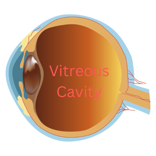
The vitreous is the substance that fills the vitreous cavity.  The vitreous cavity, or posterior chamber, is the largest compartment of the eye and is located between the lens and the retina.  The retina is the inside lining of the eye. 

Normally, the vitreous is optically clear, color-less and has the consistency of a gel.  The vitreous is composed mostly of water, but has about 5% protein. 

The vitreous is essential for normal embryologic development of the eye.  Once we are born, we are not sure of any true functions of the vitreous although many believe it is essential in oxygenation of the tissues of eye, such as the lens.

The vitreous may absorb energy.  For instance when you are engaged in sports, running, or suffer a head injury, the vitreous may be absorbing shock waves.  This is unproven.

What are floaters?

The vitreous is never regenerated.  While the fluid component does renew (aqueous humor), the proteins do not.  With time and normal aging, the proteins can denature or breakdown and the vitreous becomes more water-like.  

The thinner, more fluid, vitreous means that the vitreous changes from a thicker gel to a more watery substance.  The proteins can more easily coalesce and clump together casting shadows on the retina…more commonly known as floaters. 

Floaters can also be a result of retinal disease, blood or inflammation.  Floaters from any cause are indistinguishable from one another (i.e. you can’t tell the difference between floaters due to blood vs. inflammation).  Only your eye doctor can tell the difference after a thorough dilated examination of your eye.

It is recommended that you alert your eye care professional should you ever experience a sudden increase in floaters.  New floaters could be a sign of a potentially blinding condition. 

Treatment of Floaters

In general, the best treatment for floaters is to treat the underlying disease.  Persistent floaters can be bothersome.  Removal via vitrectomy may be the only solution for clearing the floaters from your vision.

Some doctors may offer Nd:Yag laser for the treatment of floaters.  There are limitations to this type of treatment and not all retina specialists view this treatment equally. 

If you would like to schedule an appointment, please call us (877) 245.2020.
Nader Moinfar, M.D., M.P.H.
Retina Specialist
Orlando, FL

Vitrectomy for Retinal Detachment Repair

This is the most common way to repair most retinal detachments and has faster healing times.

Vitrectomy for retinal detachment repair is an alternative method to scleral buckle or pneumatic retinopexy.

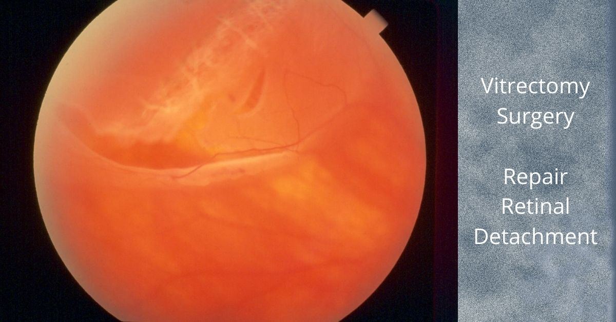
A vitrectomy is a common surgical repair for a detached retina. The surgery removes the vitreous gel from the eye, pushes the detached retina back into place, and seals any retinal tears in the retina. Then gas is injected into the eye to help keep the retina in place and prevent any fluid from leaking from the repaired tears and into the subretinal space.

The Surgery

A vitrectomy is usually performed using local anesthetic. A heavy sedative is administered and then Lidocaine is injected around the eye to numb it. If patients prefer not to be awake during the procedure they can be given intravenous sedation or a general anesthesia.

The surgery consists of making three small needle-sized incisions (about the width of an eyelash) into the white part of the eye (sclera). Through one incision a constant flow of fluid is passed into the eye. The second incision is used to insert a fiber-optic light to provide illumination of the eye, and the third is for any instruments that are used, including a “vitrector” for the removal of the vitreous.

A vitrector is an oscillating microscopic cutter that slowly removes the vitreous. Once the vitreous is removed, a freezing instrument or a laser is used to seal any tears in the retina.

The vitreous will be replaced with a gas. In time the gas will dissipate and be replaced by the eye’s own fluids (aqueous humor). The vitreous gel does not grow back, but the eye can function with just the fluid that replaces the vitreous. Depending on the type of gas used and its concentration, it can take from three to ten weeks to fully dissipate.

Most retina specialists will perform vitrectomy for retinal detachment repair in an outpatient setting. The surgery time ranges from 20 to 40 minutes, but varies greatly.

Head positioning

Head positioning after surgery is necessary to keep the gas bubble in the proper place. If the retina detached at the bottom, the only way to get the bubble to press against the place of detachment is to place the head down. The bubble walls off the damaged area while it heals and prevents any fluid from flowing through the damaged area of the retina and into the subretinal space.

The gas bubble wiur vision while it is in place, but as it dissipates a line will form across your vision where the newly forming fluid (aqueous humor) is gradually replacing the bubble. The line will move lower each day and the field of vision will get larger.

If you have to position your head down there are pillows, chairs, and mirrors that allow you to see around the room while in a face-down position.

Gas bubble precautions

As long as the gas bubble remains in your eye, you must not fly in an aircraft. The reduced pressure in the cabin will cause the gas bubble to expand. This would be extremely painful and could lead to loss of sight. If you must have general anesthesia for any surgical procedure while the gas bubble is in your eye, you must inform your anesthesiologist so you will not be given nitrous oxide, which would cause a dangerous rise in eye pressure in the eye with the gas bubble.

Safety

Vitrectomy surgery was first developed in 1970 and since then many improvements in instrumentation and technique have dramatically improved the safety of the procedure. Severe complications are rare and the surgical success rate is over 90%.  

Nader Moinfar, M.D., M.P.H.
Retina Specialist
Orlando, FL

If you would like to schedule an appointment, please call us (877) 245.2020.
Jon Doe