What is the Retina?

The retina is the light sensitive tissue that translates light into electrical signals for the brain. That’s how we see.

The retina is a highly specialized layer of tissue that lines the inside of the eye. As part of the central nervous system, it serves as a crucial component of the visual system. Its primary function is to convert light into electrical signals that can be translated to “vision” by our brain.

It consists of several layers of cells, each with a specific role in the process of vision. The outermost layer contains the photoreceptor cells, known as rods and cones. Rods are responsible for vision in low-light conditions and are highly sensitive to light, while cones are responsible for color vision and visual acuity in brighter light conditions. Cones are further classified into three types: red, green, and blue cones, allowing us to perceive a wide range of colors.

Converts Light to Electrical Signals

When light enters the eye, it passes through the cornea and lens, which help focus the light onto the retina. The rods and cones in the retina absorb the light and convert it into electrical signals. This process involves the activation of light-sensitive pigments within the photoreceptor cells, triggering a cascade of chemical reactions that generate electrical signals.

The electrical signals produced by the rods and cones are then processed by several layers of cells within the retina. Bipolar cells receive the signals from the photoreceptor cells and transmit them to ganglion cells, which are the final layer of cells in the retina. The ganglion cells collect the signals and their axons form the optic nerve, which carries the visual information from the retina to the brain.

Highly Specialized Cells Sharpen Vision

Within the retina, there are also other types of cells, such as horizontal cells and amacrine cells, which help refine and modulate the visual signals. These cells play a role in enhancing contrast, adjusting sensitivity to light, and facilitating communication between different parts of the retina.

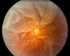
The central region of the retina is known as the macula, which contains a high concentration of cones. At the center of the macula is a small depression called the fovea, where visual acuity is highest. The fovea contains a high density of cones, allowing for detailed and sharp central vision.

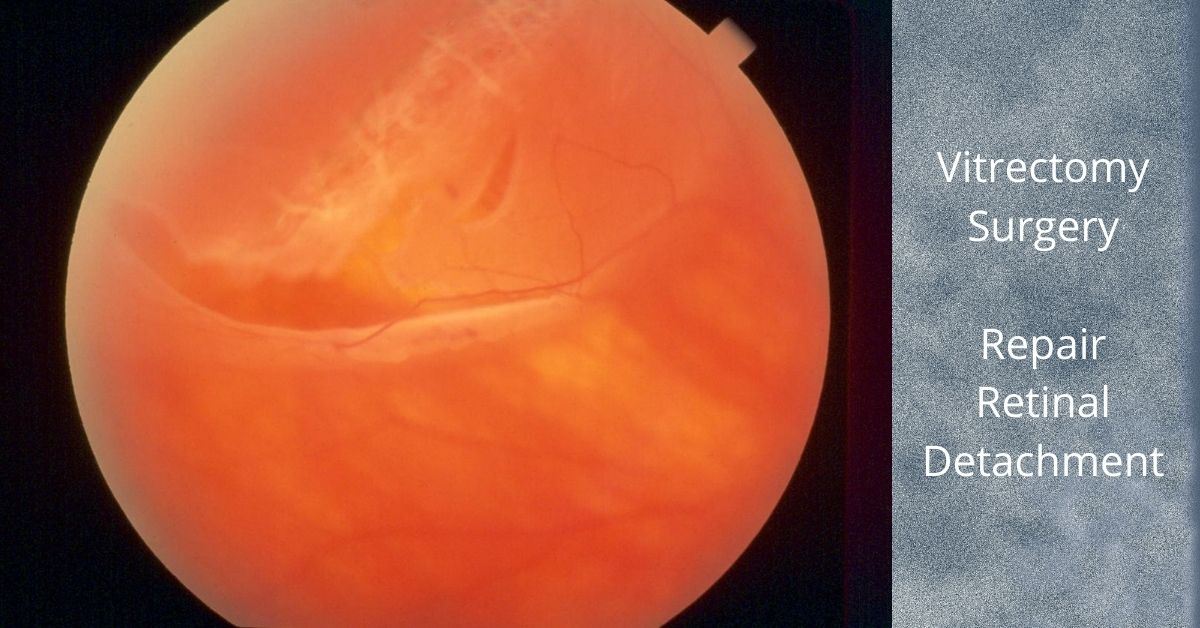
For us to see, all layers of the retina must be healthy, maintain normal blood supply and remain attached.

If you would like to schedule an appointment, please call us (877) 245.2020.

Nader Moinfar, M.D., M.P.H.
Retina Specialist
Orlando, FL

Vitrectomy for Retinal Detachment Repair

This is the most common way to repair most retinal detachments and has faster healing times.

Vitrectomy for retinal detachment repair is an alternative method to scleral buckle or pneumatic retinopexy.

A vitrectomy is a common surgical repair for a detached retina. The surgery removes the vitreous gel from the eye, pushes the detached retina back into place, and seals any retinal tears in the retina. Then gas is injected into the eye to help keep the retina in place and prevent any fluid from leaking from the repaired tears and into the subretinal space.

The Surgery

A vitrectomy is usually performed using local anesthetic. A heavy sedative is administered and then Lidocaine is injected around the eye to numb it. If patients prefer not to be awake during the procedure they can be given intravenous sedation or a general anesthesia.

The surgery consists of making three small needle-sized incisions (about the width of an eyelash) into the white part of the eye (sclera). Through one incision a constant flow of fluid is passed into the eye. The second incision is used to insert a fiber-optic light to provide illumination of the eye, and the third is for any instruments that are used, including a “vitrector” for the removal of the vitreous.

A vitrector is an oscillating microscopic cutter that slowly removes the vitreous. Once the vitreous is removed, a freezing instrument or a laser is used to seal any tears in the retina.

The vitreous will be replaced with a gas. In time the gas will dissipate and be replaced by the eye’s own fluids (aqueous humor). The vitreous gel does not grow back, but the eye can function with just the fluid that replaces the vitreous. Depending on the type of gas used and its concentration, it can take from three to ten weeks to fully dissipate.

Most retina specialists will perform vitrectomy for retinal detachment repair in an outpatient setting. The surgery time ranges from 20 to 40 minutes, but varies greatly.

Head positioning

Head positioning after surgery is necessary to keep the gas bubble in the proper place. If the retina detached at the bottom, the only way to get the bubble to press against the place of detachment is to place the head down. The bubble walls off the damaged area while it heals and prevents any fluid from flowing through the damaged area of the retina and into the subretinal space.

The gas bubble wiur vision while it is in place, but as it dissipates a line will form across your vision where the newly forming fluid (aqueous humor) is gradually replacing the bubble. The line will move lower each day and the field of vision will get larger.

If you have to position your head down there are pillows, chairs, and mirrors that allow you to see around the room while in a face-down position.

Gas bubble precautions

As long as the gas bubble remains in your eye, you must not fly in an aircraft. The reduced pressure in the cabin will cause the gas bubble to expand. This would be extremely painful and could lead to loss of sight. If you must have general anesthesia for any surgical procedure while the gas bubble is in your eye, you must inform your anesthesiologist so you will not be given nitrous oxide, which would cause a dangerous rise in eye pressure in the eye with the gas bubble.

Safety

Vitrectomy surgery was first developed in 1970 and since then many improvements in instrumentation and technique have dramatically improved the safety of the procedure. Severe complications are rare and the surgical success rate is over 90%.  

Nader Moinfar, M.D., M.P.H.
Retina Specialist
Orlando, FL

If you would like to schedule an appointment, please call us (877) 245.2020.

What is Scleral Buckle Eye Surgery

Scleral buckle eye surgery has been around for generations and is still a mainstay of retinal detachment surgery.

Scleral buckle eye surgery to repair a retinal detachment has been in use since the early 1950s and it remains an essential technique for reattaching the retina.

The scleral buckle, which is usually made of silicone semi-hard plastic, supports the retina from outside the eye. It is placed under the eye muscles and usually encircles the sclera or white part of the eye and eliminates the tugging that is detaching the retina. 

The buckle is placed behind the eyelids and cannot be seen. It is sewn to the sclera (the actual white part of the eye) and it presses the walls of the eye inward, taking pressure off the retina and allowing it to reattach to the eye’s interior wall. The buckle usually remains in place permanently.

Scleral buckling is sometimes a stand-alone treatment for the repair of a retinal detachment and sometimes it is used in combination with a vitrectomy.

A laser or a sub-zero probe (cryopexy) may also be used to seal off the areas of the retina that were torn to prevent further fluid leakage and pressure on the retina.

Side effects of a scleral buckle

The possible complications or side effects of scleral buckling surgery include:

  • Increased myopia (nearsightedness) caused by the increased length of the eye when it is buckled
  • Increase in astigmatism also caused by the buckle changing the eye’s shape
  • Double vision is uncommon but can occur because the eye’s muscles were manipulated when placing the buckle under the eye muscles.
  • Pain after the procedure which can usually be controlled with acetaminophen or ibuprofen and will subside in a few days.

 What causes retinal detachment?

The most common type of retinal detachment, that accounts for 90% of all retinal detachments,occurs when a small tear or hole appears in the retina that allows the vitreous gel of the eye to seep behind the retina where it pushed the retina away from the back of the eye. 

Tears in the retina are caused by vitreous gel, which contains millions of fibers, that becomes “sticky” and tugs at the retina making minute tears in it. Retinal holes develop in areas where the retina has thinned. Retinal holes are typically smaller than tears and have a much lower risk for causing a retinal detachment.

Recovery after scleral buckling eye surgery

The recovery time varies, but ranges from two-to-eight weeks. An eye patch may be required for a few days after surgery and antibiotic eye drops may be prescribed to prevent infection. Your eye may have redness, tenderness, and swelling for a few weeks after surgery.

If you would like to schedule an appointment, please call us (877) 245.2020.

Nader Moinfar, M.D., M.P.H.
Retina Specialist
Orlando, FL

Gas Bubbles and Retina Surgery

Not all retina surgery requires a gas bubble, but here’s when it is necessary for your retina specialist to use a gas.

Not all retina surgery requires injection of a gas bubble.

To safely work on the retina, your retina specialist must be able to safely access the inner layer of the eye. A vitrectomy, the procedure to remove the vitreous, allows the retina specialist specialist to work on the retina. 

A vitrectomy (removal of the vitreous humor gel) is done for the following conditions:

The Gas Bubble

A tamponade in medical terms is something used to close or block a wound or body cavity to stop bleeding or fluid leakage.

Not all retina surgery requires gas to be injected into the eye. The most common use of gas is for repair of a retinal detachment or macular hole. In the case of the retinal detachment (specifically a rhegmatogenous retinal detachment which is caused by a retinal tear or retinal hole), the gas is used to block (tamponade) the migration of fluid to through the tear to underneath the retina.

In the case of a macular hole, the gas is used to allow the hole to slide closed by surface tension.

The gases commonly used are sulfur hexafluoride (SF6) and perfluoropropane (C3F8), and air. Each of the gases dissipates and is replaced by natural fluid, but the time for complete dissipation varies. Air dissipates and is replaced by natural fluid in 5 to 7 days. Sulfur hexafluoride (SF6) dissipates in 10 to 14 days, and perfluoropropane (C3F8), in 55 to 65 days.

The gas bubble blurs your vision while it is in place. As the bubble dissipates you will see a line across your vision where the gas meets the newly forming fluid which is gradually replacing the bubble. The line will move lower each day and your field of vision will get larger as the natural fluid continues to replace the bubble.

As long as any of the gas bubble remains in your eye you must not fly in an airplane because the bubble can expand in the reduced pressure of the cabin causing severe pain and possible loss of sight.

Head Position

Your surgeon will ask you to position yourself in a specific way during healing and that position is dependent on what part of the retina was repaired.

In cases of macular holes, a face down position is common. Head positioning for retinal detachments depends upon the location of the retinal tear(s).

Gas Bubble Injected Last

Most retina surgery is outpatient surgery and can be done under local anesthetic and mild sedation. The sedation is given by IV and is used for anxiety relief and to put you into a relaxed and sleepy state, known as a “twilight state”. In that state you are conscious and still able to hear and follow simple instructions from your surgeon.

General anesthesia can be used for patients with dementia, severe anxiety, or young children.

Most retina surgeries take less than an hour and some less than 30 minutes. The gas bubble is injected as one of the last steps of the surgery.

Recovery

You will have to wear an eye patch for a day or two following surgery. Recovery time depends on the procedure you had, but is generally two to four weeks. An exception is the repair of a complete retinal detachment which could take several months to heal and for vision to stabilize.

If you would like to schedule an appointment, please call us (877) 245.2020.

Nader Moinfar, M.D., M.P.H.
Retina Specialist
Orlando, FL

21st Century Retina Surgery

Most retina surgery is performed on an outpatient basis, and is very similar to cataract surgery.

Retina surgery is complex and requires exacting precision within the microscopic space of the retina.  Recent advances in the size and precision of surgical instruments, microscopic viewing systems, and vitrectomy machines with multiple customizable controls have enabled retina repairs that were impossible just a decade ago.

In addition, these advances in technology have reduced operating times and many retinal repairs take less than an hour and can be done under a local anesthetic and a mild sedative at an outpatient surgical site.

Retina Surgery

To repair retinal holes or tears, remove scar tissue, or reattach a detached retina, the retina specialist must have access to the retina. To do that the vitreous humor gel that fills the eye cavity is removed in a procedure called a vitrectomy. Once the vitreous humor gel is out of the way, the surgeon makes the needed repairs to the retina. 

An EKG and blood pressure, and oxygen sensors are placed to monitor vital signs and sedation is administered through an IV. The sedation puts you in a twilight state in which you are very sleepy and relaxed yet still able to hear your surgeon and respond to simple instructions.

Eye drops are used to numb the eye. Once the eye is numb an eyelid holder is placed to prevent blinking during the surgical procedure. The eyelid holder is not uncomfortable because while your eyes are numb you will not have the sensation of needing to blink.

The Gas Bubble

After your retina is repaired a gas or air bubble is used as a tamponade to prevent the fluid that naturally exudes from inflamed tissues from reaching your retina. The gas bubble walls off and protects the repaired retina while it heals. The gas bubble gradually dissipates and is replaced by natural aqueous fluid.

There are two medical gases that are commonly used. One of them dissipates in 10 to 14 days, whereas the other takes 55 to 65 days to dissipate. If air is used as the bubble, it will absorb within 5 to 7 days.

Depending on your repair, your retina specialist will choose the appropriate medical gas with the correct absorption time.  

The gas bubble makes vision extremely out of focus while it covers the entire vitreous chamber. While it dissipates a line will appear in your vision where the bubble is gradually being replaced by aqueous humor. The line will move further down, and your field of vision will grow larger day by day.

You will be instructed not to fly in an airplane as long as any of the gas bubble remains in your eye. The bubble can expand in the reduced pressure of an airplane cabin causing severe pain and possible loss of sight.

Head Position

You may be asked to keep you head face down during your recovery to keep the gas bubble in the correct position. You can get face down pillows, chairs, and mirrors to help you see things around you while your head is face down. Your insurance might cover the cost of some of the face down recovery equipment.

After Your Retina Surgery

Your eye will be patched after surgery and you will be asked to wear the patch for a day or two following surgery. Recovery time depends on the procedure you had, but ranges from two weeks to several months for a repaired detached retina. 

If you would like to schedule an appointment, please call us (877) 245.2020.

Nader Moinfar, M.D., M.P.H.
Retina Specialist
Orlando, FL

Lattice Degeneration

Many people are told they have “thinning of the retina,” but it’s real name is lattice degeneration and it could cause a retinal detachment.

Lattice degeneration is a fairly common, but abnormal, “thinning of the retina” that can cause a retinal detachment.  This type of degeneration affects the peripheral retina, and for reasons poorly understood, increases the chances of developing retinal tears or retinal holes that can lead to a retinal detachment

Lattice degeneration may be found in 8-10% of the population and seems to be found more frequently in families with this peripheral degeneration, myopia and a few rare diseases:

“Lattice” has characteristic findings, but only the presence of retinal holes is concerning.  Not all areas of lattice develop retinal holes.  When found, your retina specialist may feel treatment is necessary to prophylax against a retinal detachment.

Symptoms of Lattice Degeneration

There are no symptoms, but the condition may predispose you to retinal tears or retinal detachment, hence, patients should be on the lookout for symptoms of retinal tears or detachment including:

  • Flashes
  • Floaters
  • Blurred vision
  • Progressive loss of peripheral vision

Patients experiencing any of these symptoms should alert their eye doctor. 

Treatment of Lattice Degeneration

The majority of patients never require treatment and can be safely monitored.  There is absolutely no threat to the vision if a tear or retinal detachment do not ensue.  

The chance of developing a retinal detachment is quite small.  Still, it is recommended that patients with this peripheral thinning be monitored regularly.  Though progression is unlikely, retinal holes can develop over time.  Retinal holes develop slowly and gradually, whereas, a retinal tear can happen suddenly.

Retinal holes from lattice degeneration are treated and managed exactly the same way as treating a retinal tear.  Not all retinal holes from lattice degeneration require treatment. 

Laser treatment is the preferred treatment, however, cryotherapy and endolaser photocoagulation with vitrectomy are viable options as well.

In short…

In summary, lattice degeneration is usually a benign “thinning” of the retina that increases your chances of developing a retinal detachment over your lifetime.  Tell your doctor if you develop sudden flashes and/or floaters or if you experience loss of your peripheral vision. 

If you would like to schedule an appointment, please call us (877) 245.2020.

Nader Moinfar, M.D., M.P.H.
Retina Specialist
Orlando, FL

Symptoms of a PVD

These symptoms may prevent a retinal detachment and here’s why.

PVD stands for “posterior vitreous detachment.” It’s a normal event that increases your risk for developing a retinal tear which can lead to a retinal detachment. Everyone will eventually get a PVD.

New flashes and new floaters should be reported to your eye doctor or retina specialist. Not all floaters are benign. Only a full dilated exam will determine if your new symptoms are related to a posterior vitreous detachment, retinal tear or something else.

Symptoms of Posterior Vitreous Detachment

The most common symptoms of a PVD:

  • Flashes
  • Floaters
  • Nothing (Asymptomatic, no flashes or floaters)

There are two ways to stimulate the retina: normal light and physical manipulation. Usually, light enters the eye and is captured by the rods and cones of the retina which eventually produce vision. The other mechanism occurs when you rub your eyes so hard you actually see patterns in your vision. This rubbing, or physical manipulation, also produces “light.”

After a PVD has occurred, only the back, or posterior portion, of the vitreous separates from the retina. The anterior portion remains fixed to the surface of the retina. As the newly separated vitreous cleaves from the retinal surface in the back, it can move to and fro in the eye. This transmits energy to the portion of the vitreous still attached to the retina in the anterior part of the eye…causing flashes.

What are Floaters?

Floaters can be a result of cellular debris from underneath the retina in cases of a retinal tear, blood or just opacities which have formed in the otherwise clear vitreous gel.

Not all posterior vitreous detachments cause flashes or floaters. Thus, follow up is always recommended so your retina specialist can look for asymptomatic retinal tears.

What if I have a Retinal Tear?

First of all new flashes and floaters should be reported to your eye doctor. In the case of a PVD, it’s important to look for a retinal tear. A retinal tear is most likely to occur during the first 6 weeks after a PVD has occurred.

The goal of examination is to find a retinal tear before it turns into a retinal detachment. Retinal tears can usually be treated with laser whereas a retinal detachment requires surgery and there is a chance of lost vision.

If you would like to schedule an appointment, please call us (877) 245.2020.

Nader Moinfar, M.D., M.P.H.
Retina Specialist
Orlando, FL

Laser Eye Surgery for the Retina

Lasers are commonly used in all of ophthalmology and are useful tools to treat disease of the cornea, glaucoma, cataracts and various retinal diseases.

Because the cornea is transparent to light, the cornea allows the laser energy enter the eye and treat the inner parts of the eye such as the retina.

Lasers for the Cornea

Reshaping the cornea allows many patients to have refractive surgery. Modern refractive surgery is called LASIK. The precision of the laser allows the surgeon to accurately resculp the curvature of the cornea to match the prescription otherwise needed by wearing glasses or contact lenses. An eximer laser is commonly used to perform LASIK.

Laser for Glaucoma

Laser surgery for glaucoma has been long been a treatment option to reduce intraocular pressure. Reducing the internal eye pressure is the mainstay of most treatments for glaucoma. Argon, diode or Neodymium:YAG lasers can be used for glaucoma laser surgery.

Cataract Laser Surgery

There are two ways lasers can assist the eye surgeon to treat cataracts and so-called after cataracts. Laser assisted cataract surgery allows the eye surgeon to more accurately perform several steps of the cataract surgery, but not the entire surgery. A femto-second laser is used for laser-assisted cataract surgery.

After initial cataract surgery, the intraocular lens commonly becomes cloudy. This cloudiness can be improved with a Nd:Yag laser.

Retina Laser Eye Surgery

Lasers are used by a retina specialist to treat 3 different types of problems:

A retinal tear can happen to anyone. Retinal tears can cause a retinal detachment which requires surgery and is vision threatening. Using an argon or diode laser, laser burns surround the retinal tear to prevent a retinal detachment from occurring from that specific tear. In essence, the retinal burn, “seals” the tear, preventing retinal detachment.

Certain types of retinal vascular occlusions (RVO) can cause swelling, aka edema, in the retina. Similar types of swelling are commonly treated in patients with diabetes. The laser acts to decrease swelling by either reducing the rate of fluid accumulation or increasing the rate of absorption.

Advanced retinal diseases, called retinopathy, can be treated with laser to stabilize the eye and prevent vision loss. Proliferative diabetic retinopathy is often treated with laser or intravitreal injections. Retinopathy can also be caused by types of vascular occlusions.

If you would like to schedule an appointment, please call us (877) 245.2020.

Nader Moinfar, M.D., M.P.H.
Retina Specialist
Orlando, FL

Flashes and Floaters

Here’s what you need to know about those pesky floaters and why they could blind you.

Eye “floaters” are deposits or debris within your eye’s vitreous humor. Whether you see specks, wispy threads, strings, or cobweb shapes that drift back and forth when you move your eyes, you are seeing floaters. More precisely, you are seeing the shadows that the floaters cast on your retinas. Floaters can refract light and that temporarily interferes with your vision until the floaters drift into a different position.  

Harmless Floaters?

Eye floaters move around, come and go, and eventually settle away from the line of sight after a few weeks. They can be annoying and distracting, but most of the time they are harmless and are not a cause for alarm, however, any sudden onset of new floaters should be examined.

Floaters are suspended in the vitreous humor which fills about 80 percent of the eye. The most common cause of floaters is shrinkage of the vitreous humor. The vitreous humor is 98 to 99% water and the remaining 1 to 2% is a mixture of collagen, proteins, salts, and sugars. Despite the water-to-collagen ratio, the vitreous humor has a firm jelly-like consistency.

Posterior Vitreous Detachment

As we age the vitreous humor shrinks and liquefies and develops a watery center. Portions of the collagen and protein mixture become stringy fibers and these floating stringy collagen and protein fibers are the floaters. This is called a PVD (posterior vitreous detachment).

Sometimes the small floating vitreous fibers pull on retinal cells and that mechanically stimulates the retina cells causing the sensation of flashes of light. This too is not unusual. Floaters and flashes occur at the same time and are mostly harmless annoyances. You have experienced mechanical stimulation of retinal cells if you’ve ever been hit in the eye and afterwards “see stars”.  

Treatments

In rare instances floaters can be so closely packed and large or so numerous that they significantly affect vision. In those cases, floaters can be treated with a surgical vitrectomy or a laser vitrectomy.  A surgical vitrectomy removes the vitreous humor and replaces it with saline or a bubble made of gas or silicone oil. A laser vitrectomy breaks apart large floaters and vaporizes them.

Floaters and flashes of light that require immediate attention

A sudden onset of numerous floaters, frequent flashes of light, a dark shadow that covers part of your side vision, or eyes that hurt can be symptoms of a tear in your retina and could indicate that your retina is detaching. A detached retina is an emergency and immediate treatment can save your sight. 

If you want to make an appointment, please call us (877) 245.2020.

Nader Moinfar, M.D., M.P.H.
Retina Specialist
Orlando, FL

Treatment of a Retinal Detachment

Retinal Detachment | Nader Moinfar MD | Lake Mary Orlando| Retina Specialist
A total retinal detachment.

A retinal detachment usually when there is pulling on the retina, which causes a tear to occur. Causes of retinal tears are multiple, and include trauma, genetic predisposition, myopia (near-sightedness), to name a few.

A retinal tear should be evaluated by a trained retina specialist, and can usually, and successfully, be simply treated in the office.

A retinal detachment is a more complicated problem, and a well- trained retina specialist can offer you different ways of treating this potentially blinding condition.

In some cases, an office-based procedure called a pneumatic retinopexy can be offered; with appropriate selection of the patient, success rates of up to 80% can be achieved. In the office, a gas bubble is injected into the eye; and either the same day, or maybe a few days laser, laser or cryotherapy is applied. Your retina specialist should discuss with you his or her experience level with this procedure, success rates, and experience levels with the other options, that may be available.

Another method to treat retinal detachments, as seen under the video section of NaderMoinfarMD.com, is a procedure called a vitrectomy. Generally speaking, the vitreous, which is the clear jelly of the eye, is the cause of a retinal tear and traction. By removing the vitreous, one removes the underlying cause. A vitrectomy for a retinal detachment must be careful and completely done, to ensure the greatest chance of success. A seen in my videos, the vitreous is removed, and following a procedure termed an air-fluid exchange, laser is applied around the tears, and finally, the space is filled with a gas bubble.

Lastly, your retina specialist may advise alone, or in combination with a vitrectomy, a procedure termed a scleral buckle; this, too, can be viewed on my video section. With this procedure, a very thin band of silicone is secured on the outside, white part of the eye, helping to relieve the internal traction on the retina; cryotherapy, and a gas bubble, are also used.

With almost all cases of retinal detachments, you will be required to keep your head and body in a certain configuration for a short time; this allows the gas inside your eye to push up against the detachment, allowing it to heal, or “stick’ down. Please note that when the gas bubble is in the eye, you should not travel by air, go up to certain altitudes, and must avoid certain types of general anesthetic inhalation agents if having surgery. Your retina specialist should provide you with a green wrist band to remind you, and others, of this precaution.

If you want to make an appointment, please call us (877) 245.2020.

Nader Moinfar, M.D.
Retina Specialist
Orlando, FL

Jon Doe