Vitreomacular traction (VMT) is a complication that arises from a normal process. That normal process is posterior vitreous detachment (PVD) and it happens to everyone as they age. By about age 40 or 50 the vitreous gel that fills the eye begins to change. It begins to shrink and lose fluid and strands of the gel can drift through the eye. These strands can be seen as dark strings or spots that float around in your field of view and are called floaters.
The vitreous gel eventually completely separates from the retina. This is perfectly normal and happens to most people by the age of 70, that is, a PVD is a completely normal event.
Problems arise only if the vitreous gel is strongly attached to the retina, specifically at the macula. If that is the case then when the vitreous gel shrinks it can pull on the retina.
That sticking process is called vitreomacular traction and is very similar to an epiretinal membrane.
The pulling and tugging on the center of the retina where the macula is located can damage the macula and cause vision loss if left untreated.
In healthy eyes, VMT is not common. Certain eye conditions or diseases put people at a higher risk of developing VMT. Those conditions and diseases include:
- High myopia which is extreme nearsightedness
- Age-related macular degeneration which is a breakdown of the tissues in the back of the eye
- Diabetic eye disease which affects the blood vessels in the back of the eye
- Retinal vein occlusion which is a blockage of veins in the retina
Symptoms
The most common symptoms of VMT include:
- Distorted vision that makes straight lines appear wavy, blurry or have blank spots
- Seeing lots of flashes of light in your vision
- Seeing objects as smaller than their actual size
It is important to see an ophthalmologist for an evaluation when you first notice any of these symptoms.
Diagnosis
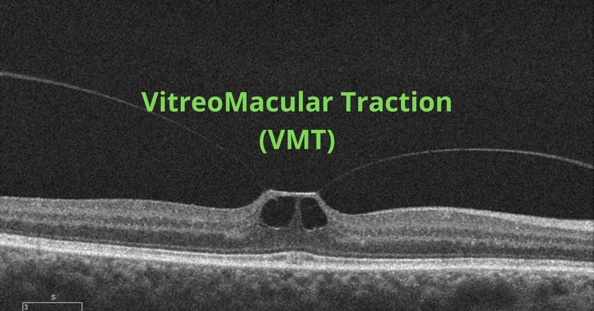
To diagnose vitreomacular traction (VMT) your ophthalmologist will use tests such as optical coherence tomography (OCT) which uses light waves to take pictures of the various layers of the retina and will show any damage to the macula.
Fluorescein angiography may also be used. This is an imaging test to view how well the blood is circulating inside the retina and to find any macula swelling. It uses a medical imaging dye injected into the arm that circulates inside the retina while a special camera photographs the progress of the dye as it moves through the blood vessels in the eye.
An ultrasound scan may also be used so your ophthalmologist can get a better view of the location of the sticking point between the vitreous and the macula.
Treatment
Some cases do not require treatment and will resolve on their own, but you will be asked to monitor your vision at home with a grid of lines to make sure the VMT does not progress. If the lines on the grid begin to appear wavy or have missing areas then you will most likely require treatment.
Surgery to remove the vitreous and replace it with a saline solution may be needed to prevent macular holes, puckers or macular swelling from developing or worsening.
Some people are candidates for medication treatments. A medication that dissolves the proteins that link the vitreous to the macula can be injected into the eye. Usually only one injection is needed.
Prognosis
Most patients with VMT maintain good visual acuity in the affected eye even if treatment is required.
If you would like to schedule an appointment, please call us (877) 245.2020.