Drug Delivery for Eye Disease

Eye drops are great for only certain eye diseases. There are other routes used to treat your retina.

There are different routes of drug delivery to treat your eye disease.

Not all parts of the eye can be reached by the same route. Eye drops and ointments applied by patients directly to the eye is the route to get to the cornea, conjunctiva, and sclera, but getting to the retina in the back of the eye requires a different route—an injection.

Intravitreal Injections

Only an intravitreal injection will reach the retina because the retina is part of the brain and is protected by the blood-brain barrier.

Intravitreal means that the injection is into the vitreous of the eye. The vitreous is a clear, colorless, jelly-like substance that fills the space between the lens and the retina. Intravitreal injections of anti-VEGF are placed in the vitreous near the retina to treat wet macular degeneration and diabetic retinopathy.

The injections are done in a doctor’s office and the eye is numbed before the injection. They are a safe and effective way to prevent further vision loss.

Eye Drops

Eye drops are the preferred route of drug delivery to treat eye diseases in the front part of the eye. They are commonly used to treat eye infections, allergies, inflammation, glaucoma, and to provide lubrication for dry eyes.

Eye drops come in solutions and suspensions. In a solution the particle sizes are very small and they completely dissolve in the solvent mixture making a solution clear. Suspensions consist of larger particles that are suspended in a solvent. The larger particles will settle to the bottom so suspensions must be shaken before use to re-suspend the therapeutic particles.

If you are applying more than one drop to an eye, allow the first drop to absorb completely before applying a second drop so the medication doesn’t end up running down your cheek.

Medicated Contact Lenses

For some corneal infections a special type of contact lens or a collagen shield is soaked in antibiotics and placed on the eye.

Ointments

Although ointments are greasy and somewhat difficult to apply they are the preferred eye medication for use on infants and very small children because they stay in the eye longer than eye drops. Ointments are also used for nighttime applications of eye medication so the ointment can cover the eye while you sleep.

Sustained Release Drug Systems

These are biodegradable, implanted devices that supply a slow, steady release of a drug over an extended time to maintain the therapeutic level of the drug to the target area. It is new technology and is available for the treatment of glaucoma, vascular occlusions and diabetic retinopathy.

If you would like to schedule an appointment, please call us (877) 245.2020.
Nader Moinfar, M.D., M.P.H.
Retina Specialist
Orlando, FL

Why Injections Not Drops

Here’s why eye drops don’t work for treating diseases of the retina.

The blood brain barrier blocks most medications from reaching the retina. You may wonder why there isn’t a pill, or an eye drop that treats these retinal diseases and why the only effective treatment is a shot in the eye, an intraocular injection.

Intraocular injections are common and effective ways to treat wet macular degeneration, diabetic retinopathy and retinal vascular occlusions.

Pills, eye drops, and even IV solutions cannot penetrate to the retina because it is protected by the blood-brain barrier. Direct injection circumvents this barrier.

The eye is the only part of the brain that can be viewed directly. And that is done when an eye doctor uses an ophthalmoscope and shines a bright light into your eye and can view the innermost layers of the eye—the retina, and the optic nerve.

Blood Brain Barrier

The blood-brain barrier (BBB) is a protective layer of endothelial cells that protects the brain from any pathogens or toxins that may be circulating in the blood supply, but it also restricts entry of large-molecule medications and 98% of all small-molecule drugs. So, the only way to get therapeutics to the brain or to the deep structures of the eye is to go through or behind the BBB.

The BBB allows water, some gasses, and fat-soluble molecules to dissolve in the cell membranes and cross its boundary. Alcohol, caffeine, and nicotine easily cross the BBB because they are lipophilic (attracted to fat) and mix easily with the fat in the BBB to gain entry. Oxygen and anesthesia are gasses that can pass through the BBB.

As part of the brain, the BBB prevents therapeutics that have been ingested and are circulating in the blood to gain entry to the eyes. The medication will travel through the body, but almost none of it will reach the eyes.

Leaky Blood Vessels

Wet macular degeneration is caused when blood vessels behind the retina grow abnormally and leak blood and fluid that distorts vision. If left untreated the leaking blood vessels will cause irreversible damage to the photoreceptors that create central vision.

A Shot in the Eye

The only way to get enough medication directly to the retina to stop the growth of the leaky blood vessels is to use an intravitreal injection. An intravitreal injection means that the medication is injected into the vitreous (jelly-like fluid) of the eye where it will diffuse to the retina.

In addition, injecting anti-vascular endothelial growth factor (anti-VEGF) directly into the eye where the leaky vessels are growing, prevents the anti-VEGF from circulating in your blood supply and possibly adversely affecting systemic blood vessels as it moves along to the eye.

The intravitreal injections are painless because the eye is numbed, and the injections can be done in your doctor’s office. They are a safe, and effective way to prevent leaky blood vessel damage to your retina.

Coming Advances in Treatment

An implanted reservoir that delivers sustained-release anti-VEGF directly to the retina is in the experimental phase. And the field of nanotechnology is in the preliminary research stages of developing nanoparticles as a potential way to transfer medication in eye drops across the BBB.

If you would like to schedule an appointment, please call us (877) 245.2020.

Nader Moinfar, M.D., M.P.H.
Retina Specialist
Orlando, FL

Retinal Vascular Occlusions (RVO)

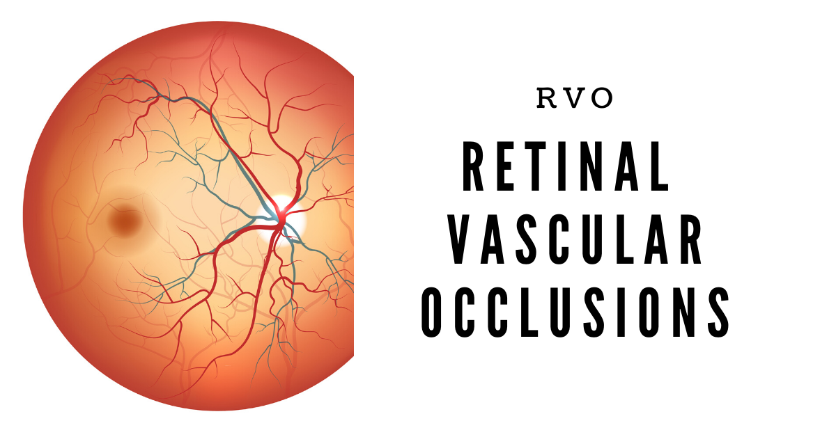
Retinal vascular occlusions cause painless loss of vision. Here’s a short review of the 4 different types.

There are four types of retinal vascular occlusions (RVO).  All have similar symptoms of acute (sudden) and painless loss of vision.  

In general, vein obstructions occur when the vessel is compressed from the outside of the blood vessel whereas artery obstructions occur when cholesterol plaque (or other substances) travel through the blood stream and become lodged in a blood vessel.

Vein and artery occlusions have characteristic appearances and can be easily diagnosed by a retina specialist.

Artery occlusions are more likely to be associated with coronary artery disease and stroke.

The four types of retinal vascular occlusions include:

  • Branch retinal vein occlusion (BRVO)
  • Central retinal vein occlusion (CRVO)
  • Branch retinal artery occlusion (BRAO)
  • Central retinal artery occlusion (CRAO)

Branch Retinal Vein Occlusion

Compared to a central vein occlusion, this is a “partial” occlusion of the retinal vein.  Usually branch occlusions involve about 50% or less of the retina. If the macula becomes involved, there is usually moderate vision loss.  On occasion, the macula is not involved and vision remains normal. 

Treatment of BRVO includes laser surgery, injections of anti-VEGF and/or intraocular steroids including Ozurdex, a sustained release device which allows continuous treatment for up to 4-6 months.

Central Retinal Vein Occlusion

The entire retinal vein is occluded.  Severe vision loss is normally experienced.  Unlike the branch vein occlusion, the macula is always involved.

Treatment of CRVO may include laser, anti-VEGF injections or sustained release steroids.  Vision usually does not improve as well as the branch retinal vein occlusion.

Branch Retinal Artery Occlusions

Branch artery occlusions do not involve the entire retina and, similar to venous occlusions, less than 50% of the retina is involved.  If the macular area is involved, central vision is decreased. 

Branch artery occlusions do cause retinal edema, but the vision loss is usually more permanent though there is resolution of the retinal swelling.

Both branch and central artery occlusions are caused by an embolus:  usually a cholesterol plaque carried through the bloodstream and becomes lodged in a retinal artery. 

Central Retinal Artery Occlusion

As with the branch artery occlusion, an embolus lodges in the central artery thus cutting off blood flow to the entire retina.  Vision loss is severe and usually does not respond well to treatment.

Historically, many different methods have been described to dislodge the embolus, but none with resounding success.

Patients with artery occlusions should be assessed for the risk of cardiovascular disease and stroke.  It’s important that your retina specialist or eye doctor coordinate care with your primary care physician. 

If you would like to schedule an appointment, please call us (877) 245.2020.
Nader Moinfar, M.D., M.P.H.
Retina Specialist
Orlando, FL

Jon Doe