Floaters from Vitreous Hemorrhage

Vitreous hemorrhage from various causes can cause floaters.

Vitreous hemorrhage can cause floaters in the eye.  Bleeding inside the eye is called vitreous hemorrhage.  Symptoms of this type of bleeding range from a showere of floaters to incomplete darkness (lots of blood).

Floaters due to blood can not be distinguished by other types of floaters based upon appearance.  Often floaters from hemorrhage have appeared suddenly and don’t necessarily last as long as chronic (long-lasting) floaters, but they can.

Causes of Vitreous Hemorrhage

Any bleeding into the vitreous is called vitreous hemorrhage.  Hence, there are a variety of causes of bleeding into the vitreous;

  1. Retinal tears
  2. Retinal Detachments
  3. Trauma
  4. Diseases of the retina (eg. proliferative diabetic retinopathy)
  5. Complications of retinal vascular disorders
  6. Posterior Vitreous Detachment

Treatments

For all intents and purposes, blood in the vitreous does not cause long-lasting damage. It is most important to establish the underlying cause of the bleeding and treat that disorder whenever possible. 

For dense vitreous hemorrhages, it may indeed be prudent to consider a vitrectomy to remove the blood to restore vision, but more importantly, and to allow the retina specialist to perform a thorough examination of the eye with the blood removed.

Retinal Tears and Bleeding

As an example, a retinal tear can cause a vitreous hemorrhage if the tear rips across a retinal blood vessel and causes bleeding.  There may be too much blood to actually visualize a retinal tear.  Keep in mind, retinal tears can lead to retinal detachments.

Retinal detachments can be potentially blinding and, in cases of all retinal tears, we would like to treat the tear before a retinal detachment develops.

Vitreous Hemorrhage Can Resolve

Blood in the vitreous usually absorbs and can clear on with a vitrectomy.  There is no specific timing of this and it can take weeks or even months.  Sometimes, blood does not clear on its own and vitrectomy may be necessary. 

If you would like to schedule an appointment, please call us (877) 245.2020.
Nader Moinfar, M.D., M.P.H.
Retina Specialist
Orlando, FL

Treatment of Diabetic Macular Edema (DME)

Diabetic macular edema is a common complication of diabetic retinopathy where fluid accumulates in the macula and blurs the vision.

Diabetic macular edema (DME) is a condition characterized by the accumulation of fluid in the macula, the central part of the retina responsible for sharp, detailed vision. It is a common complication of diabetic retinopathy, a disease affecting the blood vessels of the retina. The management of diabetic macular edema (DME) involves various treatment approaches aimed at reducing macular edema and preserving vision.

Anti-VEGF Injections – Treatment of Diabetic Macular Edema

One of the primary treatment options for DME is intravitreal injections of anti-vascular endothelial growth factor (anti-VEGF) medications. Drugs like ranibizumab, bevacizumab, and aflibercept are commonly used to inhibit the abnormal growth of blood vessels and reduce leakage, thus decreasing macular edema. These injections are typically administered on a monthly or as-needed basis, with the aim of improving visual acuity and reducing central retinal thickness.

Steroids for Diabetic Macular Edema

Another treatment modality for DME is the use of corticosteroids. Intravitreal injections of triamcinolone acetonide or dexamethasone implants can help suppress inflammation and reduce fluid accumulation in the macula. These steroid injections are usually performed at longer intervals compared to anti-VEGF agents due to their sustained-release properties.

In some cases, laser photocoagulation may be employed to treat DME. Focal laser treatment can be used to seal leaking blood vessels and reduce fluid accumulation. However, this approach is typically reserved for cases where the edema is away from the central macula to avoid damaging central vision.

New Treatments

Recently, a new class of medications called integrin receptor antagonists has shown promise in the treatment of DME. These drugs, such as pegpleranib and abicipar, target specific proteins involved in the pathogenesis of macular edema and are administered via intravitreal injections.

In addition to these specific treatments, managing the underlying diabetes is crucial in preventing and managing DME. Maintaining optimal blood sugar control, blood pressure, and cholesterol levels can help reduce the risk and progression of DME.

Regular eye examinations and early detection of DME are essential for timely intervention and optimal outcomes. Treatment options should be individualized based on the severity of the edema, visual impairment, and patient characteristics. By employing a combination of these treatment modalities and a comprehensive approach to diabetes management, healthcare professionals can improve visual outcomes and enhance the quality of life for individuals living with DME.

If you would like to schedule an appointment, please call us (877) 245.2020.

Nader Moinfar, M.D., M.P.H.
Retina Specialist
Orlando, FL

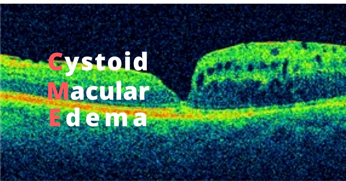
Cystoid Macular Edema

This type of macular swelling is very common and even occurs after cataract surgery.

What is Cystoid Macular Edema?

Cystoid macular edema, aka CME, affects the functional center of the retina, the macula.  It is another term for macular edema where multiple cyst-like areas of swelling develop.

There are multiple causes of CME to include:

  • Retinal Vascular Occlusions (RVO)
  • Intraocular inflammation (uveitis or iritis)
  • Diabetic retinopathy
  • Uncomplicated cataract surgery

Symptoms of Cystoid Macular Edema

The main symptom of CME is blurry vision.  There may also be some symptoms of distortion, poor color perception and minification, but most patients note blurry vision.

Because this is a condition of the macula, only central vision is affected.  The peripheral or side vision is not involved.

Blurry vision can occur from a variety of eye conditions and is not a specific symptom of cystoid macular edema. 

CME after Cataract Surgery

About 1-3% of patients will develop cystoid macular edema following cataract surgery.  This can occur several weeks following uncomplicated cataract surgery.  Before modern phacoemulsification techniques evolved, CME developed in as many as 10% of patients.

For reasons unknown, fluid starts to accumulate in the macula.  Patients are often frustrated because the vision following surgery was excellent, only to get worse. 

Treatment and Prognosis

CME following cataract surgery is usually self limited, that is, there is full restoration of vision and function after several weeks of treatment.  The condition does not usually recur, but there is increased risk of developing CME in the fellow eye.

Treatment for CME following cataract surgery often involves topical anti-inflammatory eye drops.  Steroid drops are often quite effective.  In more complicated cases, injections of steroid around the eye may be helpful

Because CME can develop from other situations other than cataract surgery, treatments and prognosis vary widely.  For instance, cystoid macular edema following a branch retinal vein occlusion may involve anti-VEGF injections or depot steroids.  

Cystoid macular edema can also be caused by certain glaucoma drops or following complicated cataract surgery.  Surgery may be necessary to treat the macular swelling due to complications of cataract surgery. 

Consultation with a retinal specialist may be needed.

Diabetic Retinopathy: Your Risk Factors

If you have diabetes, what’s your risk of developing diabetic retinopathy?

What’s your risk of developing diabetic retinopathy?

Diabetic retinopathy is the most common diabetic eye disease and a leading cause of blindness in American adults.  

Anyone with type 1 or type 2 diabetes can develop diabetic retinopathy, but your risk of developing diabetic retinopathy increases the longer you have diabetes.

According to the National Eye Institute, 2 in 5 Americans with diabetes have some stage of diabetic retinopathy. But you can lower your risk of developing diabetic retinopathy by controlling your blood sugar levels.

Women who develop gestational diabetes are at high risk for developing diabetic retinopathy. If you are diabetic and are pregnant, have a comprehensive dilated eye exam as soon as possible. Ask your doctor if you will need additional eye exams during your pregnancy.

Major Risk Factors:

  • Long-duration of diabetes (10+ years)
  • Poor blood sugar and blood pressure control

Additional Risk Factors:

  • Becoming pregnant if you are diabetic or developing gestational diabetes during a pregnancy
  • Having diabetes and being African-American, Hispanic, or Native American
  • High cholesterol
  • Smoking

Complications 

Retinal detachment. Diabetic retinopathy causes an abnormal growth of blood vessels which can also produce scar tissue. The scar tissue can pull the retina away from the back of the eye. This may cause spots that float in your vision, flashes of light, or a loss of vision. 

Glaucoma. The abnormal blood vessels may grow in the front part of your eye and interfere with the normal flow of fluid out of the eye and cause excessive eye pressure. Over time this pressure can damage the optic nerve that carries images from your eyes to your brain.

Vitreous hemorrhage. The abnormal blood vessels can also bleed into the clear, jelly-like substance that fills the center of your eye. If there is only a small amount of bleeding you will see a few dark spots (floaters). If the bleeding is severe it can fill the vitreous and block your vision.

Unless your retina is damaged, the blood will clear from your eye in a few weeks. The blood often clears from the eye within a few weeks or months. Unless your retina is damaged, your vision will return to normal.

Blindness. Left untreated, diabetic retinopathy can lead to complete vision loss.

Preventions

Control your blood sugar. You can help control your blood sugar by making healthy food choices and getting at least 150 minutes of moderate exercise each week. That amount of exercise could be accomplished by taking a brisk 30-minute walk 5 times a week. Make sure you take any diabetes medication as directed.

Monitor your blood sugar. Ask your doctor how frequently you should test your blood sugar. You may need to do so several times a day.

Get an A1c test. The A1c test will show you and your doctor your average blood sugar level for a two-to-three-month period. This monitoring will help you learn if your diabetes has been under good control and help you and your doctor make beneficial changes to your diet or medications if needed.

Keep your blood pressure and cholesterol under control. Eating a healthy diet, regular exercise and losing weight can help. You may also need some medications.

Get a comprehensive dilated eye exam at least once a year. Regularly monitoring the health of your eyes allows your eye doctor to start treatment before in the early stage of diabetic retinopathy any complications develop.

Don’t smoke. Smoking increases your risk of developing diabetic retinopathy and other complications of diabetes.  

If you would like to schedule an appointment, please call us (877) 245.2020.

Nader Moinfar, M.D., M.P.H.
Retina Specialist
Orlando, FL

Diabetes: You Can Take Charge

November is Diabetes Awareness. Newly diagnosed with diabetes or know someone? Share this article.

Diabetes is a serious life-long condition, but there’s a lot you can do to protect your health. If you have been newly diagnosed with type 2 diabetes or prediabetes, this article offers you tips for managing your diabetes.

Overtime complications of diabetes caused by uncontrolled blood sugar can develop in the eyes (diabetic retinopathy), the kidneys (diabetic nephropathy), and the feet (diabetic neuropathy). Those three areas all receive their blood supply from tiny blood vessels called microvasculature that are susceptible to damage from high blood sugar levels.  

However, when blood sugar is well controlled the rate of complications falls. A 1% decrease in the A1c test that is maintained decreases your risk of microvascular complications by about 37%.

Taking care of your diabetes or prediabetes right after diagnosis will pay off in your long-term health.

Managing Your Type 2 Diabetes

The following steps will help keep your blood sugar levels closer to normal and maintaining close-to-normal blood sugar levels can delay or prevent diabetic complications:

  • Weight loss and maintenance of the weight loss
  • Healthy diet
  • Regular exercise
  • Blood sugar monitoring
  • Using diabetes medications as prescribed

Watch your diet

There is no “diabetes diet”, but making efforts each day to take the following steps will help put you on the right track for lowering your blood sugar levels:

  • Eat more fruits and vegetables
  • Eat more foods high in fiber
  • Eat fewer refined carbohydrates, such as white bread, white rice, pastries, pancakes, and crackers
  • Eat fewer saturated fats, such as fatty cuts of beef, pork, and lamb, processed meats, butter and full-fat dairy
  • Eat fewer overall calories

You don’t have to entirely cut out foods high in saturated fats, but you should limit your consumption. Check labels and choose foods that have more unsaturated than saturated fats. 

Fatty fish such as salmon, tuna, sardines, mackerel, herring, and trout are good sources of long-chain omega-3 fatty acids and are a healthy addition to your diet.   

Get Moving

Fitness is an important aspect of managing type 2 diabetes. It can be walking, bike riding, or swimming laps, but the key is to find activities you enjoy and will keep doing. 

Just 30 minutes a day of exercise will, in time, decrease your blood sugar levels, increase your cardiovascular fitness, strengthen your bones, reduce excess body fat, and boost muscle power. Walking will also improve your mood especially if you walk in an area with some greenery or water.

Monitor Your Glucose         

Frequently check your blood sugar levels. Doing so can help you see which foods or activities trigger blood sugar highs or lows. Keep your food and activity consistent and test your blood sugar and look for patterns over 2 to 3 days. The goal is to find the insulin dose that keeps your blood sugar levels stable without causing a low.

Have regular A1c tests that will assess your average blood sugar level for the past 2-to-3 months.

Your blood sugar monitoring schedule is individual to you and should be set according to your needs and under the supervision of your doctor.

Connect with a Diabetes Care and Education Specialist

Diabetes care and education specialists are certified healthcare professionals with specialized knowledge in diabetes self-management and care. They will help you chart and then monitor the best diabetes healthcare course for your situation.

Visits to accredited diabetes education specialists are covered by most insurance plans and by Medicare Part B. You can locate an accredited diabetes education program by clicking here.

If you would like to schedule an appointment, please call us (877) 245.2020.

Nader Moinfar, M.D., M.P.H.
Retina Specialist
Orlando, FL

Saving Your Vision: Diabetic Retinopathy

November is National Diabetes Awareness month.

If you have diabetes, take a few minutes to learn about diabetic retinopathy and how you can take steps to help keep your eyes healthy.

Diabetic retinopathy is the most common cause of vision impairment and blindness among working-age adults in the U.S. It occurs when high blood glucose levels damage the blood vessels at the back of the eye causing them to become weak and leaky and damage the retina.

If not found and treated early, diabetic retinopathy can cause permanent vision loss. That’s why it is important to manage your diabetes and stay alert to the early signs and symptoms of diabetic retinopathy.

Types of Diabetic Retinopathy

There are two main types of diabetic retinopathy—NPDR (non-proliferative diabetic retinopathy) and PDR (proliferative diabetic retinopathy). The “proliferative” refers to new blood vessel growth, so in non-proliferative, there is not yet new blood vessel growth.   

NPDR is the early stage of diabetic retinopathy. At this stage, high glucose levels have weakened the blood vessels and, in time, may cause bulges in the weakened vessels (microaneurysms) that may leak fluid into the retina and the leakage may swell the macula or some blood vessels may begin to close. So this is an ongoing disease cycle that can be slowed or stopped if caught early.

PDR is the more advanced stage of diabetic retinopathy. The NPDR has weakened the blood vessels or closed them, so new blood vessels have grown in an attempt to supply oxygen to the retina, but these new vessels are fragile, and they leak and damage the retina. 

PDR can lead to vitreous hemorrhage, traction retinal detachment, or neovascular glaucoma.

Stay Alert

Anyone with type 1 or type 2 diabetes, or women who have gestational diabetes during pregnancy, can develop diabetic retinopathy. The risk increases when blood sugar, blood pressure, and cholesterol levels are not under control.

In the early stages diabetic retinopathy may not have symptoms, so it’s important to have a comprehensive dilated eye exam once a year. That way diabetic retinopathy can be diagnosed and treated before symptoms appear.   

Symptoms that indicate diabetic retinopathy has progressed are:

  • Blurry vision
  • Vision that goes from blurry to clear
  • Increased number of floaters
  • Poor night vision
  • Spots or blank areas in your central vision
  • Colors appear faded

Diabetic Retinopathy Treatment

The best treatment happens before you develop symptoms. Strict control of your blood sugar will significantly reduce your risk of vision loss from diabetic retinopathy. If you have high blood pressure and kidney problems, make sure those are kept under control too.

Injections in the eye of anti-VEGF medication can stop the growth of new blood vessels. VEGF stands for vascular endothelial growth factor, and it is a signal protein your body produces that stimulates new blood vessel formation, but the new vessels are weak and leaking vessels that damage your eyes, and anti-VEGF injections will stop that abnormal blood vessel growth.

Laser surgery and vitrectomy are other treatments for advanced PDR. But the most important thing to remember if you have diabetes, is that control of your blood sugar and early diagnosis and treatment of diabetic retinopathy are the best protections against vision loss.

If you would like to schedule an appointment, please call us (877) 245.2020.

Nader Moinfar, M.D., M.P.H.
Retina Specialist
Orlando, FL

Jon Doe