Articles

21st Century Retina Surgery

Most retina surgery is performed on an outpatient basis, and is very similar to cataract surgery.

Retina surgery is complex and requires exacting precision within the microscopic space of the retina.  Recent advances in the size and precision of surgical instruments, microscopic viewing systems, and vitrectomy machines with multiple customizable controls have enabled retina repairs that were impossible just a decade ago.

In addition, these advances in technology have reduced operating times and many retinal repairs take less than an hour and can be done under a local anesthetic and a mild sedative at an outpatient surgical site.

Retina Surgery

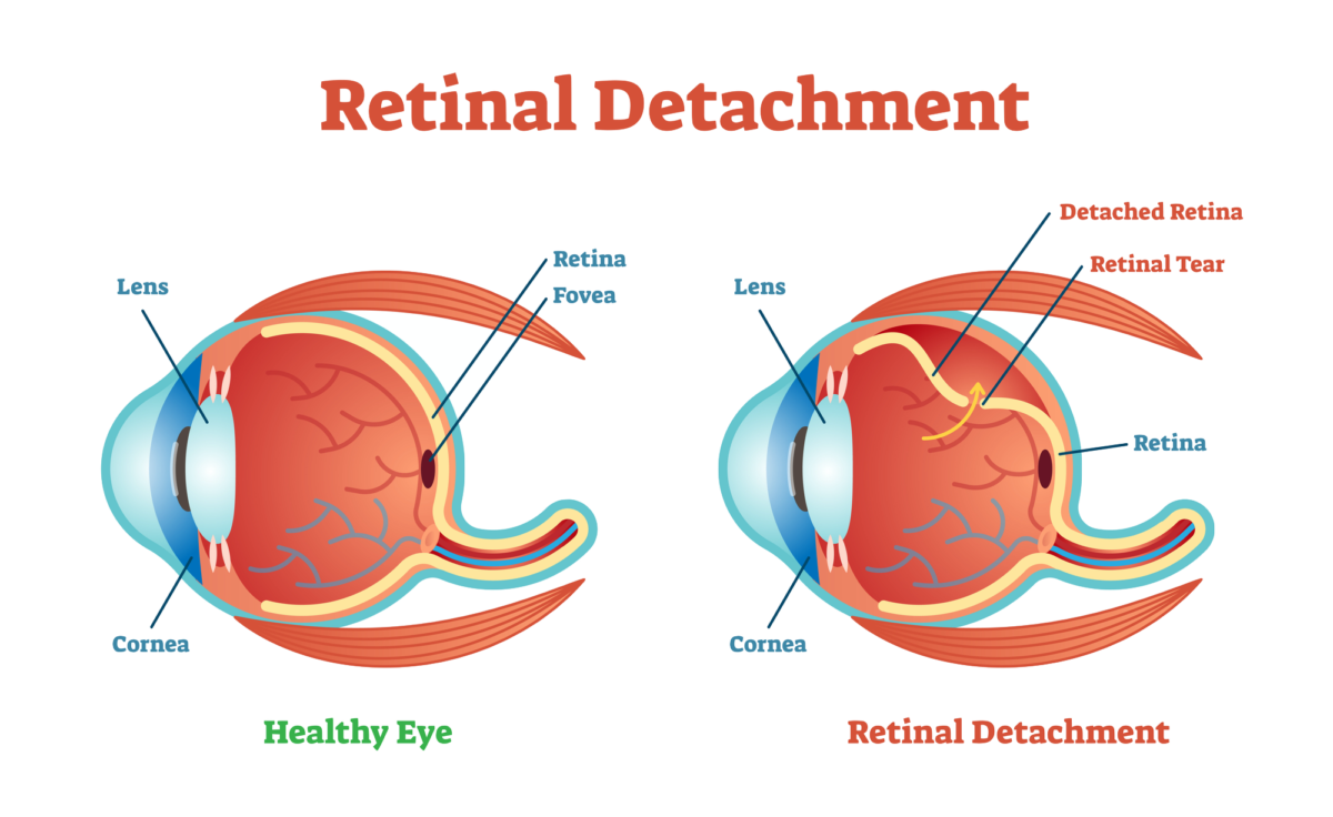
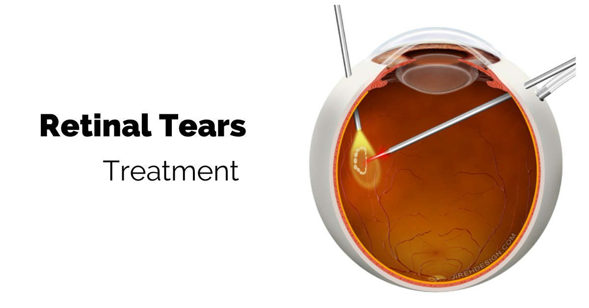
To repair retinal holes or tears, remove scar tissue, or reattach a detached retina, the retina specialist must have access to the retina. To do that the vitreous humor gel that fills the eye cavity is removed in a procedure called a vitrectomy. Once the vitreous humor gel is out of the way, the surgeon makes the needed repairs to the retina. 

An EKG and blood pressure, and oxygen sensors are placed to monitor vital signs and sedation is administered through an IV. The sedation puts you in a twilight state in which you are very sleepy and relaxed yet still able to hear your surgeon and respond to simple instructions.

Eye drops are used to numb the eye. Once the eye is numb an eyelid holder is placed to prevent blinking during the surgical procedure. The eyelid holder is not uncomfortable because while your eyes are numb you will not have the sensation of needing to blink.

The Gas Bubble

After your retina is repaired a gas or air bubble is used as a tamponade to prevent the fluid that naturally exudes from inflamed tissues from reaching your retina. The gas bubble walls off and protects the repaired retina while it heals. The gas bubble gradually dissipates and is replaced by natural aqueous fluid.

There are two medical gases that are commonly used. One of them dissipates in 10 to 14 days, whereas the other takes 55 to 65 days to dissipate. If air is used as the bubble, it will absorb within 5 to 7 days.

Depending on your repair, your retina specialist will choose the appropriate medical gas with the correct absorption time.  

The gas bubble makes vision extremely out of focus while it covers the entire vitreous chamber. While it dissipates a line will appear in your vision where the bubble is gradually being replaced by aqueous humor. The line will move further down, and your field of vision will grow larger day by day.

You will be instructed not to fly in an airplane as long as any of the gas bubble remains in your eye. The bubble can expand in the reduced pressure of an airplane cabin causing severe pain and possible loss of sight.

Head Position

You may be asked to keep you head face down during your recovery to keep the gas bubble in the correct position. You can get face down pillows, chairs, and mirrors to help you see things around you while your head is face down. Your insurance might cover the cost of some of the face down recovery equipment.

After Your Retina Surgery

Your eye will be patched after surgery and you will be asked to wear the patch for a day or two following surgery. Recovery time depends on the procedure you had, but ranges from two weeks to several months for a repaired detached retina. 

If you would like to schedule an appointment, please call us (877) 245.2020.

Nader Moinfar, M.D., M.P.H.
Retina Specialist
Orlando, FL

Drug Delivery for Eye Disease

Eye drops are great for only certain eye diseases. There are other routes used to treat your retina.

There are different routes of drug delivery to treat your eye disease.

Not all parts of the eye can be reached by the same route. Eye drops and ointments applied by patients directly to the eye is the route to get to the cornea, conjunctiva, and sclera, but getting to the retina in the back of the eye requires a different route—an injection.

Intravitreal Injections

Only an intravitreal injection will reach the retina because the retina is part of the brain and is protected by the blood-brain barrier.

Intravitreal means that the injection is into the vitreous of the eye. The vitreous is a clear, colorless, jelly-like substance that fills the space between the lens and the retina. Intravitreal injections of anti-VEGF are placed in the vitreous near the retina to treat wet macular degeneration and diabetic retinopathy.

The injections are done in a doctor’s office and the eye is numbed before the injection. They are a safe and effective way to prevent further vision loss.

Eye Drops

Eye drops are the preferred route of drug delivery to treat eye diseases in the front part of the eye. They are commonly used to treat eye infections, allergies, inflammation, glaucoma, and to provide lubrication for dry eyes.

Eye drops come in solutions and suspensions. In a solution the particle sizes are very small and they completely dissolve in the solvent mixture making a solution clear. Suspensions consist of larger particles that are suspended in a solvent. The larger particles will settle to the bottom so suspensions must be shaken before use to re-suspend the therapeutic particles.

If you are applying more than one drop to an eye, allow the first drop to absorb completely before applying a second drop so the medication doesn’t end up running down your cheek.

Medicated Contact Lenses

For some corneal infections a special type of contact lens or a collagen shield is soaked in antibiotics and placed on the eye.

Ointments

Although ointments are greasy and somewhat difficult to apply they are the preferred eye medication for use on infants and very small children because they stay in the eye longer than eye drops. Ointments are also used for nighttime applications of eye medication so the ointment can cover the eye while you sleep.

Sustained Release Drug Systems

These are biodegradable, implanted devices that supply a slow, steady release of a drug over an extended time to maintain the therapeutic level of the drug to the target area. It is new technology and is available for the treatment of glaucoma, vascular occlusions and diabetic retinopathy.

If you would like to schedule an appointment, please call us (877) 245.2020.

Nader Moinfar, M.D., M.P.H.
Retina Specialist
Orlando, FL

Why Injections Not Drops

Here’s why eye drops don’t work for treating diseases of the retina.

The blood brain barrier blocks most medications from reaching the retina. You may wonder why there isn’t a pill, or an eye drop that treats these retinal diseases and why the only effective treatment is a shot in the eye, an intraocular injection.

Intraocular injections are common and effective ways to treat wet macular degeneration, diabetic retinopathy and retinal vascular occlusions.

Pills, eye drops, and even IV solutions cannot penetrate to the retina because it is protected by the blood-brain barrier. Direct injection circumvents this barrier.

The eye is the only part of the brain that can be viewed directly. And that is done when an eye doctor uses an ophthalmoscope and shines a bright light into your eye and can view the innermost layers of the eye—the retina, and the optic nerve.

Blood Brain Barrier

The blood-brain barrier (BBB) is a protective layer of endothelial cells that protects the brain from any pathogens or toxins that may be circulating in the blood supply, but it also restricts entry of large-molecule medications and 98% of all small-molecule drugs. So, the only way to get therapeutics to the brain or to the deep structures of the eye is to go through or behind the BBB.

The BBB allows water, some gasses, and fat-soluble molecules to dissolve in the cell membranes and cross its boundary. Alcohol, caffeine, and nicotine easily cross the BBB because they are lipophilic (attracted to fat) and mix easily with the fat in the BBB to gain entry. Oxygen and anesthesia are gasses that can pass through the BBB.

As part of the brain, the BBB prevents therapeutics that have been ingested and are circulating in the blood to gain entry to the eyes. The medication will travel through the body, but almost none of it will reach the eyes.

Leaky Blood Vessels

Wet macular degeneration is caused when blood vessels behind the retina grow abnormally and leak blood and fluid that distorts vision. If left untreated the leaking blood vessels will cause irreversible damage to the photoreceptors that create central vision.

A Shot in the Eye

The only way to get enough medication directly to the retina to stop the growth of the leaky blood vessels is to use an intravitreal injection. An intravitreal injection means that the medication is injected into the vitreous (jelly-like fluid) of the eye where it will diffuse to the retina.

In addition, injecting anti-vascular endothelial growth factor (anti-VEGF) directly into the eye where the leaky vessels are growing, prevents the anti-VEGF from circulating in your blood supply and possibly adversely affecting systemic blood vessels as it moves along to the eye.

The intravitreal injections are painless because the eye is numbed, and the injections can be done in your doctor’s office. They are a safe, and effective way to prevent leaky blood vessel damage to your retina.

Coming Advances in Treatment

An implanted reservoir that delivers sustained-release anti-VEGF directly to the retina is in the experimental phase. And the field of nanotechnology is in the preliminary research stages of developing nanoparticles as a potential way to transfer medication in eye drops across the BBB.

If you would like to schedule an appointment, please call us (877) 245.2020.

Nader Moinfar, M.D., M.P.H.
Retina Specialist
Orlando, FL

Surgery for Retinal Detachment

Here’s how a retinal detachment occurs and the various surgical methods used by a retina specialist to repair this potentially blinding problem.

There is a variety of surgery to repair a retinal detachment.

Retinal detachments are potentially blinding and usually require surgery. Retina specialists are the type of eye doctor who specialize in this type of surgery.

The retina is the electrical circuitry of our eyes, and it covers about 65% of the back of the eye. It contains rod and cone photoreceptor cells that are biological transducers, which means the cells translate physical stimuli (the light reflected off objects) into electro-chemical signals that are sent to the visual cortex in the brain via the optic nerve where they create our vision.

When the retina has a tear or a partial detachment that section of the circuitry can no longer send visual signals. There is no visual input from the torn or detached area.

Retinal detachment surgery may be a medical emergency and must be treated quickly. The longer and greater the extent of the retinal detachment, the worse the visual outcome.

Symptoms of Retinal Detachment

  • Flashes of light
  • Lots of new floaters (small black specks or threads) in your vision.
  • A shadow or “curtain” descending from the top or across the side of your eye

Some retinal detachments happen gradually and are preceded by flashes of light and floaters and others detachments happen rapidly and the only symptom is the descending dark curtain blocking vision where the retina has detached.

Causes

An injury to the face or eye can detach a retina. Aging can cause changes in the gel-like vitreous material inside the eye. When the changed vitreous moves around it can tug on the retina and cause a detachment. Tumors and some diseases such as diabetes can cause retinal detachments.

A posterior vitreous detachment (PVD) is a common cause of retinal tears leading to retinal detachment.

Treatments

Pneumatic Retinopexy is the simplest fix for small retinal detachments and the procedure can be done in the office. With your eye numbed, a gas bubble is injected into your eye so that it presses against the detachment to hold it in place and then the area is sealed using a cryogenic (freezing) probe or a laser. After the procedure you must keep your head in the position recommended by your doctor for about 1 to 3 weeks.

Scleral buckling is a surgical procedure performed in the operating room.  The sclera is the white outer layer of the eye and your surgeon attaches a band made of silicone around the sclera from top to bottom. This compresses the eye and pushes the retina back into place where it will reattach to its blood supply and heal.

Vitrectomy is the removal of the vitreous gel that fills the eye. The retina specialist removes any scar tissue and seals any retinal tears and then fills the eye with saline, air, or a gas bubble. Over time your eye displaces and replaces them with its own fluid. The vitreous does not regenerate, but your eye is able to function well without it.

Retina specialists vary in the their approach to retinal detachment surgery, and often, a combination of the methods and techniques that are described above can be utilized.

Recovery

Vision will gradually improve after retina reattachment. It may take up to six months or longer to regain your best vision. An air or gas bubble injected into the eye will temporarily blur your vision until the bubble dissipates.

Less vision will be recoverable If the detachment was severe and the central portion of the retina was detached, or if treatment was delayed and the retina was without blood flow for an extended time.

Not all retinal detachments situations are the same and it is recommended you communicate with your own doctor about your prognosis.

If you would like to schedule an appointment, please call us (877) 245.2020.

Nader Moinfar, M.D., M.P.H.
Retina Specialist
Orlando, FL

Lattice Degeneration

Many people are told they have “thinning of the retina,” but it’s real name is lattice degeneration and it could cause a retinal detachment.

Lattice degeneration is a fairly common, but abnormal, “thinning of the retina” that can cause a retinal detachment.  This type of degeneration affects the peripheral retina, and for reasons poorly understood, increases the chances of developing retinal tears or retinal holes that can lead to a retinal detachment

Lattice degeneration may be found in 8-10% of the population and seems to be found more frequently in families with this peripheral degeneration, myopia and a few rare diseases:

“Lattice” has characteristic findings, but only the presence of retinal holes is concerning.  Not all areas of lattice develop retinal holes.  When found, your retina specialist may feel treatment is necessary to prophylax against a retinal detachment.

Symptoms of Lattice Degeneration

There are no symptoms, but the condition may predispose you to retinal tears or retinal detachment, hence, patients should be on the lookout for symptoms of retinal tears or detachment including:

  • Flashes
  • Floaters
  • Blurred vision
  • Progressive loss of peripheral vision

Patients experiencing any of these symptoms should alert their eye doctor. 

Treatment of Lattice Degeneration

The majority of patients never require treatment and can be safely monitored.  There is absolutely no threat to the vision if a tear or retinal detachment do not ensue.  

The chance of developing a retinal detachment is quite small.  Still, it is recommended that patients with this peripheral thinning be monitored regularly.  Though progression is unlikely, retinal holes can develop over time.  Retinal holes develop slowly and gradually, whereas, a retinal tear can happen suddenly.

Retinal holes from lattice degeneration are treated and managed exactly the same way as treating a retinal tear.  Not all retinal holes from lattice degeneration require treatment. 

Laser treatment is the preferred treatment, however, cryotherapy and endolaser photocoagulation with vitrectomy are viable options as well.

In short…

In summary, lattice degeneration is usually a benign “thinning” of the retina that increases your chances of developing a retinal detachment over your lifetime.  Tell your doctor if you develop sudden flashes and/or floaters or if you experience loss of your peripheral vision. 

If you would like to schedule an appointment, please call us (877) 245.2020.

Nader Moinfar, M.D., M.P.H.
Retina Specialist
Orlando, FL

Treatment of Retinal Tears

Here are common ways a retina specialist can treat a retinal tear to prevent a retinal detachment.

Retinal tears can lead to a retinal detachment.  A retinal detachment can cause permanent vision loss and possible blindness.

Successful treatment of a retinal tear or retinal hole can prevent a retinal detachment from occurring. 

Posterior Vitreous Detachment

Also known as a PVD, a posterior vitreous detachment is the most common cause of a retinal tear.  The most common symptoms of a PVD are sudden flashes and floaters.

Any acute flashes or floaters require a complete dilated examination of the retina looking specifically for a retinal tear.  

Retinal Holes

Retinal holes are usually not caused by a posterior vitreous detachment, trauma or other eye surgery.  Most retinal tears are associated/caused by lattice degeneration, a common “normal” finding in many near-sighted patients. 

For the purposes of this article, both retinal tears and retinal holes are treated using the same techniques. 

Laser Treatment for Retinal Tears

The preferred treatment of a retinal tear is with laser treatment.  Laser light is used to create scarring around the retinal tear to prevent fluid from migrating underneath the retina.  This does not really fix the retinal tear, but it does prevent a retinal detachment from occurring. 

Laser treatment can be performed easily in the office setting and usually does not require any aftercare.  Most retinal specialists will recheck the retina a few weeks after laser treatment. 

Laser treatment usually does not cause pain when applied.  There are no nerves in the retina, but there are some deeper nerves in certain locations of the eye that can cause “discomfort.”

Cryotherapy for Retinal Tears

Not all tears can be treated with the laser.  Cryotherapy, an older treatment, can be equally effective.

For laser treatment to succeed, your retinal specialist must be able to see the entire retinal tear.  Some tears can not be completely visualized due to:

  • Vitreous hemorrhage (blood)
  • Cataract
  • Anterior location (hard to “see” the entire retina without special techniques/instruments)
  • Intraocular implants

Cryotherapy treats retinal tears by freezing them from the outside of the eye.  Because cryotherapy requires manipulation and indentation of the eye, more tears can be treated that otherwise could not be fixed with laser. 

Vitrectomy

As a last resort, intraocular surgery called vitrectomy can be used to treat a retinal tear.  In this situation, a laser probe is introduced inside the eye at the time of surgery to treat the tear.  Using the endolaser, the tear or hole is treated from the inside of the eye.  While virtually any tear can be treated in this fashion, it does require an invasive procedure and must be performed in the operating room. 

Regardless of the modality of treatment, the results are the same.  The goal is to prevent retinal detachments from occurring. 

If you would like to schedule an appointment, please call us (877) 245.2020.

Nader Moinfar, M.D., M.P.H.
Retina Specialist
Orlando, FL

Symptoms of a PVD

These symptoms may prevent a retinal detachment and here’s why.

PVD stands for “posterior vitreous detachment.” It’s a normal event that increases your risk for developing a retinal tear which can lead to a retinal detachment. Everyone will eventually get a PVD.

New flashes and new floaters should be reported to your eye doctor or retina specialist. Not all floaters are benign. Only a full dilated exam will determine if your new symptoms are related to a posterior vitreous detachment, retinal tear or something else.

Symptoms of Posterior Vitreous Detachment

The most common symptoms of a PVD:

  • Flashes
  • Floaters
  • Nothing (Asymptomatic, no flashes or floaters)

There are two ways to stimulate the retina: normal light and physical manipulation. Usually, light enters the eye and is captured by the rods and cones of the retina which eventually produce vision. The other mechanism occurs when you rub your eyes so hard you actually see patterns in your vision. This rubbing, or physical manipulation, also produces “light.”

After a PVD has occurred, only the back, or posterior portion, of the vitreous separates from the retina. The anterior portion remains fixed to the surface of the retina. As the newly separated vitreous cleaves from the retinal surface in the back, it can move to and fro in the eye. This transmits energy to the portion of the vitreous still attached to the retina in the anterior part of the eye…causing flashes.

What are Floaters?

Floaters can be a result of cellular debris from underneath the retina in cases of a retinal tear, blood or just opacities which have formed in the otherwise clear vitreous gel.

Not all posterior vitreous detachments cause flashes or floaters. Thus, follow up is always recommended so your retina specialist can look for asymptomatic retinal tears.

What if I have a Retinal Tear?

First of all new flashes and floaters should be reported to your eye doctor. In the case of a PVD, it’s important to look for a retinal tear. A retinal tear is most likely to occur during the first 6 weeks after a PVD has occurred.

The goal of examination is to find a retinal tear before it turns into a retinal detachment. Retinal tears can usually be treated with laser whereas a retinal detachment requires surgery and there is a chance of lost vision.

If you would like to schedule an appointment, please call us (877) 245.2020.

Nader Moinfar, M.D., M.P.H.
Retina Specialist
Orlando, FL

Board Certified Eye Doctor

What does board certified really mean…and what’s a retina specialist?

I am both a board certified ophthalmologist (eye doctor) and a retina specialist. Board certification requires periodic testing of knowledge.

To become a retina specialist required additional training following my completion of ophthalmology residency.

The test to become a board certified certified ophthalmologist had nothing to do with my decision to become a retina specialist. Board certification is required to obtain hospital privileges, state licensure and, perhaps, participate in many health insurance plans.

Ophthalmology Residency

After completion of medical school, all graduates are technically an “M.D.”

Internship: In most cases, the first year after graduating from medical school is spent as an intern where we gain practical experience in various core fields of medicine such as internal medicine, general surgery or some combination of the other specialties. We do not focus on ophthalmology.

Residency: Ophthalmology residency begins after the internship. Most residency programs are 3 years long and for this period, we learn nothing but ophthalmology. Residents are essentially apprentices to the craft of ophthalmology learning from practicing ophthalmology attending physicians.

At the end of residency, many of my colleagues chose to start private practice in general ophthalmology. A general ophthalmologist usually performs cataract surgery, treats various diseases such as glaucoma, diabetic retinopathy, etc.

Fellowship Trained Retina Specialist

The period of training following residency is dedicated to sub-specialty training and is called a fellowship. To become a retina specialist, I completed an additional 2 years of training after completion of residency.

In other words, after I was qualified to become a general ophthalmologist, I took on optional training to allow me to become a retina specialist.

There are about eight or recognized sub-specialties within ophthalmology. Diseases of the retina is one of the these sub-specialty areas.

The sub-specialties in ophthalmology are:

  • Cornea/Refractive
  • Glaucoma
  • Neuro-ophthalmology
  • Ocular pathology
  • Oculoplastics
  • Pediatrics
  • Oncology
  • Uveitis
  • Retina Specialist

Most fellowship trained doctors eventually practice only their sub-specialty, that is, as a retina specialist, I only take care of patients with problems with the retina.

While I am qualified to perform cataract surgery and diagnose glaucoma, I choose not to.

Board Certified

What does this mean?

Board certification, at least in ophthalmology, is a test or certification of competency in general ophthalmology. It has nothing to do with my being a retina specialist. At this time, there is no sub-specialty board certification.

Though I am a retina specialist, my board certification reflects that I am fluent and knowledgeable in all the various areas of ophthalmology. This is true of all ophthalmologists who practice a sub-specialty.

“Board certified” simply means I know a lot about general ophthalmology.

If you would like to schedule an appointment, please call us (877) 245.2020.

Nader Moinfar, M.D., M.P.H.
Retina Specialist
Orlando, FL

What is Legal Blindness?

Legal blindness can be caused by a variety of diseases. Here’s how it is defined and links to resources.

Legal blindness is a legal definition that is used to refer to patients with vision loss who qualify for government assistance. This is not total blindness.

Legal blindness is defined as a patient’s best visual acuity (aka best corrected) in either eye is 20/200 or less or the size of the visual field (peripheral vision) is less than 20 degrees. In other words, if both eyes do not see well, the vision is no better than 20/200 in better seeing eye.

“Best corrected” refers to the use of the best corrective lens/contact lens for the patient.

A central visual acuity of 20/200 or worse means that a legally blind person must be 20 feet away from an object to see what a “normal” person can see from 200 feet away.

Loss of peripheral vision can also qualify as legal blindness. Most patients normal peripheral vision can see at least 140 degrees without turning the head. Legally blind peripheral vision is less than 20 degrees.

Causes of Legal Blindness

  • Age-related macular degeneration (ARMD) is one of the leading causes of legal blindness in Americans aged 60 and older. ARMD affects the macula (functional center of the retina) and therefore decreases central vision.
  • Cataracts will affect almost everyone by the age of 80. Cataracts blur both central and peripheral vision (though reversible with cataract surgery).
  • Diabetic retinopathy is another leading cause of blindness and affects the blood vessels in the back of the retina. Diabetic retinopathy causes either blurring of the central vision (most common), but can affect peripheral vision, too.
  • Glaucoma is a progressive disease that damages the optic nerve. Glaucoma primarily causes visual field loss and, only in the late stages, affects central vision.

NOTE: All of the disease described above have available treatments which, in most cases, can preserve vision or slow down progression if diagnosed timely.

Low Vision Aids

May low-vision aids and devices are available to assist individuals who are legally blind. Both near and distance vision can be improved with low-vision aids.

For example, desktop, stand-alone and hand-held magnifiers are available. These may be helpful for close range work such as reading or computer use. Prices vary up to several hundred dollars.

Wearable devices that magnify are also available. Mounted binoculars, wearable HD autofocus cameras with TV viewing and electronic headsets with built-in cameras can help patients with central and peripheral vision loss. Prices vary up to several thousand dollars.

Resources

There are many governmental and non-governmental resources for those who are legally blind.

For example, the American Foundation for the Blind (AFB) can assist those with low-vision. Founded in 1921, the AFB ensures that patients who are blind, legally blind or otherwise visually impaired have access to educational materails, technology and legal information.

Here are additional resources that help blind, legal blindness and visually impaired.

Low Vision Evaluation

If you feel you suffer from legal blindness or need more information, a low-vision examination is the first place to start. Eye doctors specializing in low vision can advise and educate you about the best low-vision aids for your specific visual needs.

If you would like to schedule an appointment, please call us (877) 245.2020.

Nader Moinfar, M.D., M.P.H.
Retina Specialist
Orlando, FL

Telemedicine for Retina Specialists

Ready for telemedicine? Here’s what you need to know to get prepared…if it’s available at all from your doctor.

It’s May 2020 and we are in the midst of the pandemic.  In most states, stay at home orders have been in effect for about 6 weeks and healthcare has been moving along with guidelines designed to not stress our healthcare system.

In March, the American Academy of Ophthalmology and Centers for Disease Control and Prevention made strong recommendations to ophthalmologists to reduce patient care to those with urgent and emergent conditions.

About the same time, the American Academy of Ophthalmology and CMS introduced several changes to promote the use of telemedicine as an alternative to face-to-face examinations.  The idea is to reduce COVID-19 exposure to ourselves, our staff and patients. 

What is Telemedicine?

Until the pandemic, telemedicine was probably used loosely to refer to the remote screening and management of patients.  Telemedicine has the potential to help patients in remote areas (rural communities, war zones) where healthcare is sparse.  Telemedcine has the potential to screen and triage patients, but since the pandemic, telemedicine might substitute as an office visit if you are unable or unwilling to expose yourself to others who might be infected. 

Most commonly, telemedicine uses our phones and computers to create audio-visual connections between patients and providers.

While the exact platforms (digital programs) vary from office to office, a connection providing audio and visual information is at the core of effective telemedicine.

Common platforms for telemedicine include:

  • Face Time
  • Facebook Messenger Video
  • Google Hangouts
  • Zoom
  • Go-to-Meeting 
  • Skype
  • Others

Many of us are already using these platforms for connecting with friends, family and business colleagues.  Ask your own doctor/healthprovider to see what platforms they are using.

What You Need for Your “Appointment”

In most cases, you’ll need your smart phone.  While an Internet connection and a computer may suffice, it may add technical complexity to speaking with your provider.  

In addition to having the proper digital tools, be prepared to have or do the following:

  • Give consent to the telehealth visit (minors need parental permission)
  • Current health problems and details
  • Medical history
  • List of medications
  • Knowledge of operating your device (e.g. can you take a selfie?)

Your eye doctor may be able to check your vision remotely and examine different parts of your eyes and face.  You will likely need to assist by changing positions and changing camera angles.  For ophthalmology, taking a “selfie” may be necessary.

Your provider should have a discussion with you – be prepared by having a list of questions ready before starting the visit. 

Your provider may ask you to follow up with pictures, emails or texts.  

Telemedicine for Retina Specialists


Telemedicine works better for some specialists and not as well for others.   For instance, telemedicine probably works great for psychiatry, but perhaps not as well for dentistry.

Similarly, a retina specialist may have more difficulty fully utilizing the advantages of telehealth because patients must have a dilated exam.  While there are screening devices available to avoid a dilated eye exam, these devices are available on a commercial basis and not available for home use.

In general, for a retina specialist to best examine and diagnose your retinal problem, dilating the pupils, optical coherence tomography and fluorescein angiography may need to be completed which requires a trip to the office.

Still, if you are having symptoms, such as acute loss of vision or pain, you may start your journey by calling your eye doctor to see if a telemedicine visit is right for you.

If you would like to schedule an appointment, please call us (877) 245.2020.

Nader Moinfar, M.D., M.P.H.
Retina Specialist
Orlando, FL

Jon Doe